


Introduction
Oxygen saturation monitors or pulse oximeters (SpO2) were entered into practice in the 1980s not only without randomized trials but also, and probably more importantly, without education of neonatal bedside care providers around the world (i. e.: nursing staff, respiratory therapists and also physicians). Education about some known physiological principles, such as the changing relation between O2 and hemoglobin, PaO2 and O2 saturation (O2Sat), did not accompany the routine implementation of SpO2 monitors.
Preterm infants of all gestational ages and postnatal ages receiving supplemental oxygen have been treated in many places using targets of O2Sat seen in normal healthy infants breathing room air (i. e.: "physiological" or "normal" values). This was accepted and entered into practice in many cases without full understanding of the meaning of what O2Sat really is and without considering the changing relationship between PaO2 and O2Sat and the differences in SpO2 monitors used. In the past few years, the findings in a number of studies have shown that the long-accepted "physiological" targets of O2Sat by many may in fact be too high. However a "comfort zone" exists in routine clinical practice and it is assumed that if the O2Sat is "high" and the baby appears pink, the preterm baby is "doing well". On the other hand, in many units, based on understanding of known and proven concepts, the targets of O2Sat were never 97-100 % for preterm infants receiving supplemental oxygen early in life.
Oximetry monitors have become commonplace in acute healthcare setting over the last 10-12 years. It has been said blood oxygen can now be considered a "fifth vital sign". By 1989 there were 29 manufacturers producing 45 different models of oximeters1. There are currently less manufacturers producing over 20 different models and recent technological advances have changed some important aspects of SpO2 performance.
All SpO2 monitors have some variability between the measurements with the actual arterial O2Sat value measured by a cooximeter, but true new generation SpO2 monitors are much better than all others. As importantly, monitors are affected by noise, but the different SpO2 monitors handle noise in different ways, leading to differences in real measurements, speed in which a problem is detected, alarms and false alarms, and the presence or not of "holding periods". In addition, newer technology offers measurements of perfusion index. It is our objective to describe all these factors and others in relation to SpO2 monitoring for neonates.
The reliability, accuracy and therefore clinical utility of pulse oximetry remain problematic in some types of infants under certain conditions. As it will be described in this manuscript, improved signal processing technology has substantially improved the ability of some oximeters to work reliably under conditions of poor perfusion, motion artifact and noise from other sources such as bilirubin lights.
Historically it was common teaching that if clinicians or bedside care providers were confronted with questionable oxygen saturation values, they should estimate the reliability of the monitor by correlating the pulse rate measured by the oximeter with the heart rate obtained by an electrocardiogram (ECG) monitor used simultaneously. As it will be shown later, accuracy in the recording of pulse rates is also a problem of many monitors.
Knowledge and understanding of bedside care providers
Unfortunately, due to incomplete educational programs, not all bedside caregivers are proficient in optimal clinical interpretation of the data provided by the monitors. Based on published evidence and in our own experience, most of the clinicians do feel that they have received adequate training in this topic. However, 80 % or so is able to correctly identify what a pulse oximeter measures. Of greater concern is that not more than 40-45 % of caregivers can correctly identify how a pulse oximeter works and, worse only 15-20 % have a correct understanding of the oxyhemoglobin dissociation curve, a key concept in the accurate interpretation of data provided by these monitors. Unfortunately, SpO2 monitors were put in practice without adequate educational programs. When hypothetical clinical situations are presented to providers, many make numerous errors and inadequate evaluations. It has been shown that even though a large percentage of neonatal staff feels well trained and knowledgeable about pulse oximetry, there is still a lack of knowledge of basic principles. For example, it is not clear to many caring for preterm babies how high can the PaO2 be when a preterm infant (and any human being) breathes a gas with oxygen supplementation and the SpO2 reads 97-100 %. In fact, in this situation the PaO2 cannot be predicted, and it can be "as low as" 60-70 mmHg or "as high" as 300 mmHg, or higher. Furthermore, not much understanding exists currently among care providers about how SpO2 monitors work, what are the differences in SpO2 monitors, and what are "truly" new technological and significant innovations and advances in relation to signal processing and "intelligent" sensors.
The gap between knowledge and practice is associated with morbidity and can sometimes be lethal. We offer this review with the hope of improving the understanding of some of these principles, which have not only educational implications but are also of significant clinical relevance.
The questions we will attempt to answer in this review are:
1.How do SpO2 monitors work?
2.How does the SpO2 monitor handle noise and altered signals?
3.Is the alarm a "true" alarm?
4.Does the monitor have "holding periods"?
5.Does the monitor perform well and accurately when is needed the most?
6.When the O2Sat reads > 96 % what is the PaO2 of the premature infant?
7.When the SpO2 monitor reads an O2Sat level, what is the true arterial O2Sat of the premature infant?
8.When the SpO2 monitor reads an O2Sat level, what is its variability ("error")?
9.When one monitor reads a certain O2Sat level, what does another monitor read?
10.Finally, how much do clinicians know about the differences in SpO2 monitors, as described technologically and in many clinical trials.
What is known to be risky can be avoided through education and expanding the knowledge and understanding of bedside care providers. Most would agree that finding "the best practice" is frequently not possible, but eradicating a "bad practice" known to be associated with increased risks and changing a monitor with false readings, false alarms and holding periods, is important and can be done and could only be beneficial in clinical practice for newborns.
We aim to review in detail all of the issues mentioned in this introduction and provide comprehensive responses to the questions posed before, so that the reader may answer them as factually as possible with the most current knowledge.
How do saturation monitors function?
Oxygenated hemoglobin (HbO2) and deoxygenated or reduced hemoglobin (Hb) absorb and transmit discrete wavelengths of light, for the red light around 660 nm and for the infrared light around 940 nm. This is according to a physical property unique to each molecular species called the extinction coefficient. The physics of pulse oximetry has been based on the Beer-Lambert law, which takes into consideration the extinction coefficient, the concentration and the optical path length. This is demonstrated in figure 1, which shows a relation between the extinction coefficient with the wavelength, in nanometers (nm), detecting both red and infrared light, at 600 nm and 905-940 nm, respectively.
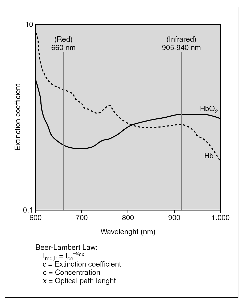
Figure 1. Relation between extinction coefficient and wavelength, in nanometers (nm), detecting both red and infra red light, at 600 nm and 905-940 nm.
The pulse oximeter sensor or probe consists of two light emitting diodes (LED), one for red light and one for infrared, and of a photodiode detector. For best performance, the LEDs and the detector must be placed on opposite sides of a translucent perfused site. In neonates, the hand or foot are commonly used. The photodiode measures three different light levels: the red light, the infra red light and also the ambient light level.
The principle that allows the transcutaneous oximeter to be an arterial or "pulse oximeter" is that it records only the transmitted light values of the added volume of arterial blood that surges through the tissues with each arterial pulse. Based on the original work of Aoyagi, it was assumed that only the arterial blood pulsates within the measuring site. This is called the pulsating arterial component [AC]. The light absorbed changes as the amount of blood in the tissue bed changes and as the relative amounts of HbO2 and Hb change. Measuring the changes in light absorption allows estimation of arterial oxygen saturation and heart rate. In addition, there is the non-moving static component [DC]. This includes tissue, bone, blood vessels, fluids, skin, and also the LED intensity, the detector response and the relatively low frequency venous blood. By dividing the pulsatile components [AC] by the non-pulsatile components [DC] for each LED, the transmission of light is normalized and calibration would not be required. As light passes through human tissue, it is absorbed in various degrees.
Thus, through spectrophotometric methods, SpO2 monitors measure HbO2 and Hb by absorption of red and infrared light. Since HbO2 and Hb absorb different amounts of red and infrared light, two wavelengths oximeters cannot measure dyshemoglobins (e. g. COHb and MetHb).
The relative proportions of HbO2 and of Hb are detected by SpO2 monitors according to the normalized ratio of light transmitted between the red and infra red lights, or R/Ir. Essentially this means that the ratio between the red and infrared wavelengths (R/Ir) at the photo detector correlates with SpO2, as shown by following the formula:

This equation needs a "look-up table" to compute the R/Ir ratio to SpO2. In brief, when the R/Ir ratio is 2.50, the SpO2 is 0 %; when it is 1.50 the SpO2 is 40 %; when the ratio reaches 0.40, the SpO2 is 100 %.
Without motion, the values of the light are transmitted through the constant portion of the arterial and venous blood, and the light transmitted through the intervening tissues is discarded (fig. 2A). However, with motion, conventional pulse oximeters do fail (fig. 2B).
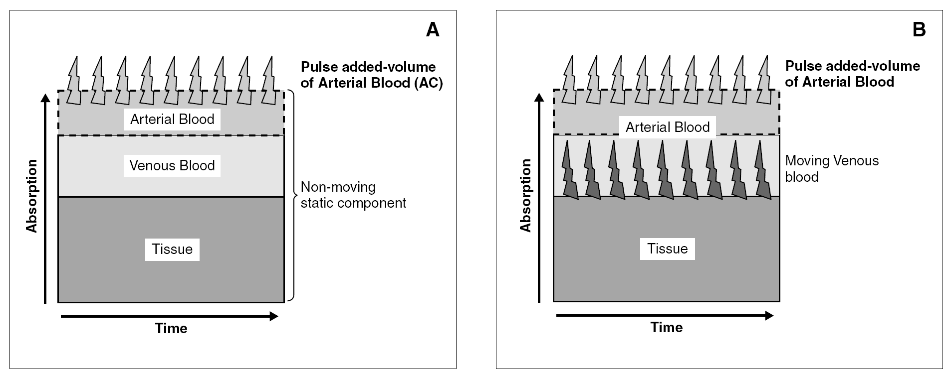
Figure 2. A) When light passes through tissues some of the light is absorbed. Without motion, the only variable light absorption is by the arterial blood (AC). B) During motion, conventional SpO2 displays falsely low value since it measures arterial AND non arterial pulsatile components.
In a few words, SpO2 estimates percent oxygen saturation of pulsatile arterial blood by measuring the absorption of light of two wavelengths and analyzing the R/Ir ratio. Instrument calibration is not required, and the probes are non-invasive and easy to apply; furthermore they do not cause significant injuries, even among the smallest newborns. A significantly high correlation of SpO2 with arterial blood oxygen saturation and partial arterial oxygen pressure (PaO2) has been reported over the normal range of blood oxygenation in healthy individuals, but there are differences among monitors. Some work very poorly under certain conditions when they are needed the most.
To measure accurately, the monitor must distinguish between the constant absorption and the pulsatile changes in absorption caused by the changing blood volume with each heart beat (fig. 2B). False readings can be due to the change in background or constant absorption, since when this changes the shape or position of the tissues through which the light passes also changes. Furthermore, when there is more motion than what can be recognized by conventional SpO2 technology, false readings are produced (fig. 2B).
Arterial oxygen saturation, oxygen content and other basic concepts related to SpO2 monitors
Arterial oxygen saturation (O2Sat) simply represents the grams of available hemoglobin concentration [Hb] which are carrying oxygen. If O2 Sat is 90 % and [Hb] is 15 g/dl, 13.5 g/dl of Hb are carrying oxygen; the other 1.5 g/dl are not. The supply and delivery of oxygen to the tissues depends on several factors and not just % O2 Sat. Among them is oxygen content of the blood. The O2 content is expressed in volume per cent (ml/dl) and is mostly dependent on % O2 Sat, [Hb] and a constant (% O2 Sat x [Hb] x 1.36). With O2 Sat of 95 % and [Hb] of 17 g/dl, there will be around 22 ml/dl of O2 in the blood. If O2 Sat falls to 80 %, the O2 content in the blood will decrease to around 19 ml/dl. Now, if O2 Sat is 80 % but [Hb] is 10 g/dl, the O2 content of the blood will be only around 11 ml/dl.
The dissolved oxygen (i. e. O2not bound to Hb) is negligible, and affects the O2 content in a minimal way even when the PaO2 is very high. For example, with maximal O2 Sat (100 %) at a PaO2 of 78 torr, increasing the PaO2 to 200 torr will have minimal effect in oxygen content and delivery but can have potential deleterious effects through superoxide anions and radical oxygen species. The essential notion is that PaO2 is needed to help saturate Hb but there is absolutely no advantage, and there are risks, to allow PaO2 to be high.
The oxygen supply and delivery depends on oxygen content and also on the flow to the tissues; this in turn is related to cardiac output (heart rate and systolic ejection volume), after load, degree of regional vasoconstriction or vasodilatation and other factors. The fundamental concept is that at maximal O2 Sat (100 %) there can be tissue hypoxia if [Hb] is low, cardiac output is decreased or local flow is altered. Similarly, with "lower" O2 Sat there can be sufficient O2 supplied and delivered. However, when the SpO2 is < 85 % there may be reasonable indication of insufficient oxygen supply to the tissues due to the associated low PaO2 levels; this would be worse if the O2 content is low due a low [Hb] (7-11 g/dl) as frequently allowed in preterm infants in many neonatal intensive care units (NICU).
The saturation reported in arterial blood gas (ABG) determinations is a calculated saturation and should not be considered in clinical practice. (Capillary samples read lower PO2 than arterial ones and therefore the O2 reported is always lower than arterial.) In ABG machines there are many changes that are not accounted for when measuring gases in blood from newborn infants. They include the changes in pH, temperature, PaCO2, 2-3DPG and fetal hemoglobin. In summary, O2 Sat reported in ABG many times has absolutely no correlation with the true arterial O2 Sat.
It is considered that at a constant [Hb], with a relatively constant pH and PaCO2, blood flow to the vital organs (brain and heart and retina) is inversely related to O2 Sat, while blood flow to the non-vital organs (gut, muscle, lung and other tissues) is directly related to the O2 Sat. Therefore, O2 Sat as measured by SpO2 can be used to estimate relative rates of organ flow and to predict changes in organ perfusion and the capacity for oxidative metabolism as oxygenation changes.
In 1986, Severinghaus2 speculated that the saturation monitor is actually a desaturation meter; that is to say that Desaturation = 100 Saturation. He more recently commented on the disagreement about the positions of sensor for the monitor readings and on the controversy regarding alarms, regulatory issues, and patent infringements. These concepts are more significant today than then.
Continuous SpO2 is now a standard of care in the NICU and has had a substantial impact on how oxygenation is managed. The confidence limits of SpO2 compared to arterial O2 Sat in a given SpO2 reading could be as high as ± 10 % when the saturation is > 70 %. When there is significant desaturation (< 70 %), the accuracy is even less. With new technology, however, the accuracy is ± 3 %. The overall accuracy of SpO2 is intrinsically limited by the use of only two wavelengths, and depends on the initial calibration population. More importantly, the empirical algorithms used to convert the signal to its real value and the quality control of the hardware together with the advances in technology, add important sources of variability between oximeters, which we as clinicians cannot ignore.
Conventional and new generation pulse oximetry
The key assumption of conventional pulse oximetry was that the only pulsating moving component was the arterial blood (fig. 2A). This was an erroneous assumption, especially during motion. During movement, or "noise," the optical path length changes dramatically and overcomes the real signal; hence motion is a physical limitation of pulse oximetry. Therefore, based on concepts that were erroneous and shown to be false, conventional pulse oximetry has many false alarms. This issue was not addressed for over a decade after the first commercial pulse oximeters were introduced. Unfortunately, the industry tried to fix the false alarm problem by introducing other problems. Products like Oxismart "masked" the false alarms when the instrument detected motion or noise. By technological methods, the industry added freezing, "holding" or zero reporting, through a decision matrix in the equipment. In summary, in many monitors when the optical path length changes dramatically and overcomes the signal during motion and "noise", the decision matrix reports a new value, reports zero or 'holds', maintaining a display from a previous period; this produces false readings and is of no use and risky in clinical conditions.
The problem therefore is that conventional pulse oximetry does not function when it is needed the most. Mostly due to motion artifact and/or low perfusion, 50-90 % of the alarms are false and over 10 % of the time these monitors fail to even measure due to low perfusion, movement or light.
Venous oxygen saturation, motion and SpO2 readings
Under no motion or noise and with strong perfusion, the venous O2 Sat at the site of sensor measurement is close to the arterial O2 Sat. During low perfusion, however, the venous O2 Sat can be 70 % lower than arterial O2 Sat. When there is movement and low perfusion, everyone can understand that there will be low amplitude of the pulsatile arterial signal which at some time or another will affect the reading of SpO2 monitors. In addition to this easily understood phenomenon, the low perfusion confuses the correct measurement because of the low venous O2 Sat that occurs (fig. 3). When the peripheral venous O2 Sat is markedly less than the arterial O2 Sat at the site of measurement, the conventional pulse oximeter now reports the average of the arterial and venous O2 Sat (fig. 3). In addition, "motion" is a big cause of error because of the movement of the venous blood, appearing like pulsating arterial blood to the pulse oximeter (fig. 2B). During motion there is low venous pressure blood "slosh" (with back and forth movement). In these situations, the AC is variable due mostly to movement of venous blood. Since conventional pulse oximetry measures both the arterial and non-arterial pulsatile components, movement of venous blood will "confuse" the monitor, which will then provide low saturation values in a false way (fig. 2B).
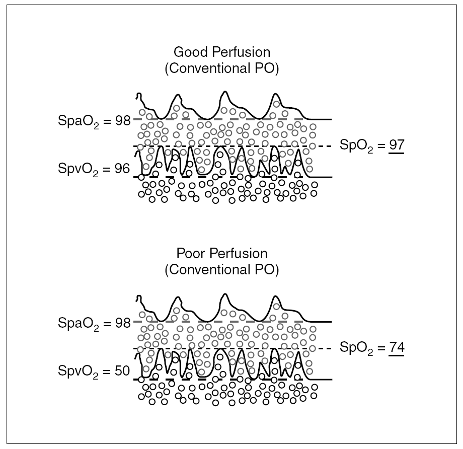
Figure 3. Influence of perfusion on the accuracy of conventional pulse oximetry during motion and how a reading will be falsely low when venous saturation is low.
The frequency and magnitude of error in SpO2 measurement is mostly influenced by: a) the venous blood saturation which is markedly lower than arterial saturation in low flow conditions; b) the magnitude of the motion and noise, and c) the arterial signal amplitude. In summary, SpO2 monitors need to work in NICU when they are needed the most (motion, low venous O2 Sat, noise and low perfusion), but this does not occur with many monitors available in the market. The fundamental concepts used by Masimo SET theory is that the light is absorbed in a very different way when there is no motion as compared to when there is motion and that measurements at low arterial signal amplitude are possible with adequate technology.
Functional and fractional saturation
There are two different types of saturation described: functional and fractional. These concepts, even though theoretical, may have an impact in the presence of dyshemoglobinemia. Functional saturation is the quantity of HbO2 expressed as a percent of hemoglobin that can transport oxygen. On the other hand, fractional saturation is the HbO2 expressed as a percent of all the hemoglobin measured (including carboxyhemoglobin and methemoglobin). Therefore, the functional saturation is:
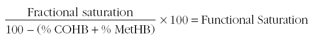
As an example to clarify the concepts above, let's assume an infant with total [Hb] of 14 g/dl. Let's assume that 7 g/dl are COHB or Met-Hb and the other 7 g/dl can transport oxygen in this extreme example. If one is making a determination by functional saturation and the grams of hemoglobin (7 g/dl) that can transport oxygen were fully saturated, the saturation reading will be 100 %. However, if we were measuring fractional saturation (and the 7 g/dl were also fully saturated), the reading will be 50 %.
Regardless whether functional or fractional saturation is used by manufacturers to calibrate their oximeters, the evaluation against fractional saturation is recommended since this is a clinically relevant variable. There is no absolute reference for oxygen saturation, although multi wavelength in vitro oximeters are accepted as the gold standard (Cooximeter).
Clinical issues related to SpO2 monitors
Clinically we need to remind ourselves that in neonatal care we use many treatments which could change perfusion, like sedation, anesthesia, dopamine and others. It is also important to remember that poorly reliable SpO2 signals occurring with hypoxemia and hypotension cannot be improved by high doses of dopamine infusion and high volume expansion. What is needed is to use monitors with adequate technology that are reliable and accurate so that motion and changes in temperature and light do not affect the true accurate reading of O2 Sat.
Pigmentation
Darkly pigmented skin can produce altered SpO2 values. It has been estimated that the degree of pigmentation in neonates, particularly preterm, is not sufficient to cause this problem. Additionally, with new pulse oximeter technology, jaundice produced by high plasma bilirubin does not interfere with SpO2 in neonates.
Ambient Light
Phototherapy can interfere with SpO2 accuracy, in the same way that there is interference caused by other sources of bright ambient light. This problem has been markedly improved by newer technologies, as described in this review.
Hemoglobin Species
Carboxy hemoglobin (HbCO). This can falsely elevate the SpO2 measurement (i. e.: SpO2 > than real SaO2), because HbCO absorbs light at the two wavelengths used in pulse oximetry to a degree similar to that of HbO2. This problem is more common in smokers, but an infant whose mother smoked heavily shortly before birth could have a relatively high HbCO concentration during the immediate postnatal period.
MET-hemoglobin (METHb). This species has a characteristic brown color that causes pulse oximeters to read closer to 85 %. However, METHb is less than one percent of total hemoglobin concentration and is not a significant concern in most clinical cases. Some drugs, however, like sulfa drugs, some local anesthetics and nitric oxide, as well as artificial blood products can generate dangerous METHb levels. In these cases SpO2, oxygen content and fractional saturation are low, but PaO2 is usually normal or high.
Fetal hemoglobin (HbF). A large proportion of HbF may erroneously decrease the arterial O2 Sat value when measured by a cooximeter, because it falsely increases the HbCO measurement, thereby falsely decreasing the percent of HbO2 measurement. HbF, however, does not have an extinction coefficient sufficiently different than that of Hb or HbO2 to measurably affect SpO2 values. In the initial period after birth, there is a high concentration of HbF and HbF has a left shifted oxygen saturation curve and a different relation with PaO2 values. For this reason HbF achieves near maximum O2 Sat levels at relatively low PaO2 values. A "clinically acceptable" O2 Sat (86 % to 93 %) maybe be associated in some cases with PaO2 levels as low as 38 to 42 torr, but in this case oxygen content and oxygen delivery may be perfectly adequate. Additionally, with high concentration of HbF an infant may be fully saturated and show a "high normal" SpO2 value (i. e. > 95 %) at lower PaO2 values than when HbF concentration decreases.
Low and high oxygen concentrations
In some cases when O2 Sat < 85 %, SpO2 has been measured to be 5-10 % higher than the real O2 Sat value. Several factors may contribute to this. Since sensor positioning is much more critical to achieving accurate SpO2 readings, sensor malpositioning may cause the SpO2 monitor to indicate only mild hypoxemia, when there is severe hypoxemia. Recently, Gerstman et al3 confirmed an important fact. He showed that conventional pulse oximeters give lower than real results when the saturation is high, and give higher than real results when the saturations are low3. This is one of the reasons why many SpO2 monitors may read 97-98 %, when the true arterial oxygen saturation measured by cooximetry is 100 %. Furthermore, in a recent clinical evaluation in children, Robertson and Hoffman4 showed that with low signal quality or during hypoxemia the new devices are not clinically equivalent to each other. Masimo SET monitor reported less data questionable than Nellcor in this study4.
Motion
An important limitation of conventional pulse oximetry has been motion artifact. As mentioned the error induced by motion can lead to unnecessary over titration of oxygen. Motion adds pulsatility to non-arterial blood components. The magnitude of error in SpO2 measurement is influenced by the venous O2 Sat, which is lower in low flow conditions, by the magnitude of the motion, the arterial signal amplitude and the technical algorithms of each SpO2 monitor. The SET technology has helped to perfect diagnostics and minimize unnecessary interventions in these conditions (see later).
Low pulse pressure
Very low perfusion states may fail to produce sufficiently large pulse amplitudes for reliable SpO2 determination. Perfusion of the infant's vascular bed between the LEDs and the sensor of the monitor's probe determines the size of the signal that is available to the pulse oximeter. As perfusion decreases, the size of the signal also decreases. When perfusion falls too low, the magnitude of the signal approaches the base system noise level of the SpO2 electronics, allowing the electronics to overcome the physiologic signal. This condition can occur in infants with heart failure, with or without shock, and also in those who have had large volume transfusions and have a high venous pressure. Dopamine may also induce a similar loss of pulse pressure by producing vasoconstriction that is associated with reverse venous pulsation. These conditions produce an extra pulse signal and are the reason why many SpO2 monitors in the market falsely estimate blood O2 Sat during such conditions.
Temperature
Low peripheral temperature with induced vasoconstriction also contributes to inaccurate SpO2 values.
Effect of altitude
Thilo et al5 studied the effect of altitude on SpO2 readings. Among healthy infants, up to three months of age, at an altitude of 1,610 meters in Denver, Colorado, the mean SpO2 was 92-93 %. The lower end of the reference range was 86 % during quiet sleep. Niermeyer et al6 studied serial SpO2 measurement from birth to four months, with healthy infants, at high altitudes at 3,100 meters. The mean SpO2 ranged from 81 ± 5 % to 91 ± 2 %, during the four month period.
Use of SpO2 monitoring in the delivery room
Heart rate and oxygenation status are essential to the delivery room assessment of newborn infants. The new generation of pulse oximeters, particularly Masimo SET, has been assessed for its use during the delivery room evaluation and management of high risk newborns7. Findings reveal a significant difference between the effectiveness of this new SET technology and also show improvement in patient outcome compared to the group with no SpO2 monitoring. This technology provides rapid acquisition and near continuous display of SpO2 values in the delivery room. This can be of significant benefit for initial respiratory care, decisions about oxygen administration and dosing and also to determine the need for more intensive procedures. Therefore, immediate or near immediate post natal SpO2 monitoring of newborns in the delivery room is now feasible and valuable.
Unapparent hypoxemia close to the time of discharge from NICU
Some clinicians prolong hospitalization because of "low" SpO2 readings. Some infants that appear ready for discharge, with no physical findings of respiratory difficulty have marginal low SpO2 readings; this can result in delaying discharge. These SpO2 readings could indeed be spurious, but at times it is difficult for clinicians to determine this carefully, particularly when arterial blood gas analyses are not readily available. The use of SpO2 may reveal sub-clinical and usually self-correcting hypoxemia that would otherwise be unknown to the clinician. The incidence of this clinically unapparent hypoxemia ranges from 20-82 %8. Some of this variation is due to differing definitions of the magnitude and duration of qualifying episodes of desaturation. The unresolved question is whether these until now unknown periods of hypoxemia should be treated and whether treating them makes a difference8.
Documentation of low and high oxygen saturation in clinical practice
Another unsolved clinical issue is whether the SpO2 saturations by oximetry monitoring are noted in the nursing notes and in the doctor's progress notes. Some evidence9 suggests that of all desaturation episodes demonstrated by pulse oximetry, only 33 % of the time will this be revealed in the nursing notes and 7 % in the doctor's progress notes. Periods of true and serious hypoxemia tend to be documented more frequently than periods of high SpO2, particularly after the FiO2 had been increased due to a "desaturation episode". This practice has to change if the main objective is to improve outcomes.
Alarms in NICUs
It has been said that as many of 86 % of ICUs alarms are false alarms. SpO2 has been particularly prone to false alarms, especially in neonatal care. Bohnhorst et al10 were able to get sensitivity in the 93-95 % range by setting the upper alarm limit of the SpO2 monitor at 95 % to avoid PaO2 > 80 torr10. They unfortunately found that this high sensitivity was associated with low specificity (26-45 %), meaning that the probability of a high alarm being false was 55-74 %.
Alarm adjustments are always made by the healthcare staff to try to minimize the number of real or false alarms at the upper end. This creates a substantial problem, because alarms are also turned off. This problem is much worse when the adequate, advanced technology described in this review is not utilized. In a report by Salyer11, when the alarm limits have an 80-90 % chance of identifying periods of hyperoxemia with older oximeters, the technicians have a hard time dealing with it because the alarm sounds too frequently. Many clinicians have become somewhat jaded about the urgency of responding to alarms. In many places, the alarm was (and still is) completely disregarded because there are so many false alarms. Alarms have become a nuisance in pulse oximetry in neonatal care and this is one of the reasons why they are very frequently turned off. This, of course, has been associated with serious, adverse consequences. The Joint Commission on Accreditation of Healthcare Organizations (JCAHO) issued a special sentinel event bulletin in February of 2002 regarding alarms. This was because of the failure of bedside care providers to apply alarms properly. We are much more hopeful now because the newer SpO2 technology gives more confidence to care providers by creating fewer false alarms and measuring saturation much more accurately.
Poets et al12 studied pulse oximetry alarm frequency. They compared Masimo SET to a conventional oximeter in non-sedated preterm infants. The median frequency of alarms per hour was 4.0 with conventional SpO2 compared to 0.3 with Masimo SET. We reported a marked decrease in alarms and significantly better handling by nursing staff when changed to Masimo SET13. It is interesting to note that noise has been shown to reduce the speed of development of neonates. Therefore, to have pulse oximeters that don't falsely alarm may have a secondary positive effect.
Since it is impossible to maintain 'perfectly steady' SpO2 and oxygen levels in blood, one should settle for a range of SpO2 values in order to decrease alarms. At the same time we should avoid drastic FiO2 changes and responding to false alarms.
Education and "Change of Culture"
An important unanswered question is why pulse oximetry was ever started in the newborn intensive care unit without adequate training and education on how to interpret the readings, how to react to the monitor display and how does technology differ between monitors.
A fundamental concept is that SpO2 was designed to measure hypoxia and has NO value in measuring hyperoxia. However, clinicians in neonatology must also know if there is hyperoxia, particularly in premature infants. Thus, oximeters need to be used with full understanding of physiology and technology. The largest problem is that pulse oximetry is used as a surrogate for arterial oxygen tension, but when the O2 Sat is $ 96 % the reliability of knowing what is the true PaO2 is lost and this can become an important problem in relation to oxygen toxicity, particularly in small premature babies. However, the process of changing the culture so that high readings are not accepted is not an easy one, as we described in our related manuscripts13,14. All of us (R.T.s, R.N.s, N.NP.s and M.D.s) must become more focused on understanding pulse oximetry and on reacting to alarms properly.
Relation between PaO2 and oxygen saturation
The relationship between PaO2 and O2 Sat has been well understood for many years. Even though there has never been convincing evidence for the rational use of supplemental oxygen in caring for preterm infants, there is clear physiological evidence and knowledge of the relationship between O2 and Hb. When an infant is breathing supplemental O2, as many are in the NICUs, we need to ask ourselves what is the PaO2 when the SpO2 is 80 %; 90 %; or 100 % or close to it? On figure 4, a summary of this relation is depicted. We do know, with close proximity, what the PaO2 is at certain O2 Sat levels in newborns. As it can be seen, within O2 Sat range of 83-93 %, the PaO2 will be between approximately 43 to 60 torr in most cases. However, we need to remind ourselves and humbly accept that when an infant is breathing supplemental oxygen we are unable to predict the PaO2 when the O2 Sat in the SpO2 monitors is > 95 %. However, at "mid and lower" SpO2 levels the ranges of PaO2 can be predicted fairly well. When the SpO2 reads 90-93 %, the PaO2 of most infants will range around 45-55 torr and will unlikely be more than 65-70 torr.
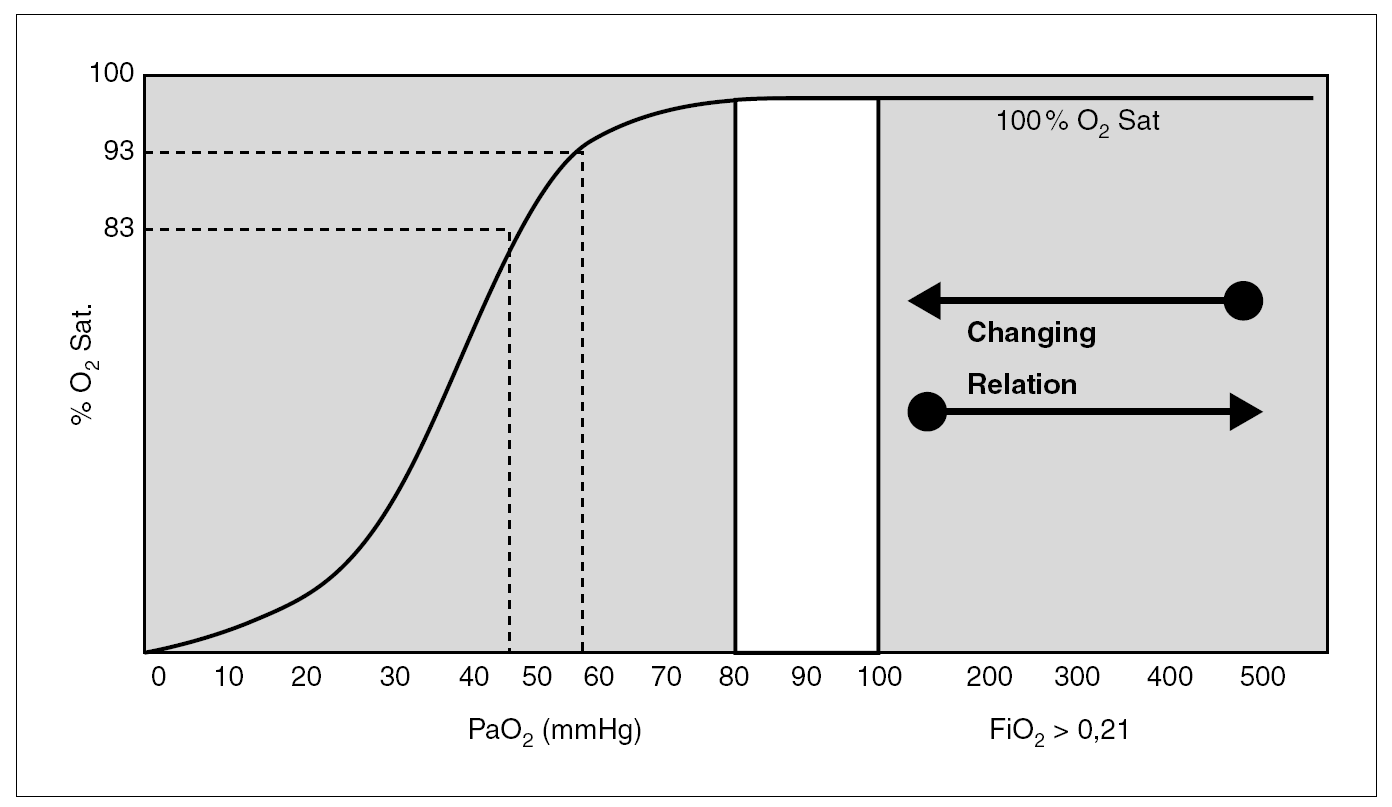
Figure 4. O2 Sat and PaO2. (Oxygen Dissociation Curve). Within a saturation range of 83-93 %, the PaO2 will be between approximately 43 to 60 torr.
When an infant with normal lungs is breathing supplemental oxygen (0.22-1.0 FiO2), if the SpO2 monitor reads 97-100 % the PaO2 could be as high as 100 torr and up to 300-500 torr. On the other hand, with FiO2 1.0 in infants with congenital heart disease, persistent pulmonary hypertension or seriously abnormal lungs, the SpO2 may be low (70-85 %) but it can also be 93-95 % and the PaO2 be around 60-70 torr.
In summary, SpO2 measurements below 80 % and above 95 % do not reliably and accurately predict PaO2. As care providers, we must remember that in addition to SpO2 inaccuracy, which lies between 0.07 to even 5 % at O2 Sat of 85 % to 100 %, SpO2 monitors do not allow accurate estimation of PaO2 when the saturations are > 95 % in infants who are breathing oxygen. At this saturation level, small SpO2 changes (1 % to 2 %) may be associated with relatively large PaO2 changes.
Normal and abnormal oxygen limits, oxygen saturation and PaO2
There is currently no definite evidence, no consensus and, therefore, no final agreement of what the optimal, ideal saturation for preterm infants is. "Normal" and acceptable saturation ranges for neonates during their acute illnesses are still to be defined. However, as clinicians, we have to make decisions continuously in this regard. High SpO2 levels for preterm infants who are being treated with oxygen are unnecessary and possibly detrimental. We need to respond to this as aggressively as we have responded to hypoxic SpO2 values, trying to avoid hypoxia, and, at the same time, avoid hyperoxia. As we describe in a separate manuscript14 our recommendations for this currently include: a) using the new Masimo SET technology from the time of birth, in order to avoid false alarms and unnecessary, inadequate clinical responses; b) from birth and during the first several weeks of life maintain SpO2 levels that do not exceed 93 % or a maximum of 95 % in premature infants breathing oxygen; c) setting the high alarm limits at these values; d) not turning alarms off. The lower level at which alarm limits should be set is still questionable at this time and we would like to stress the caveat that the number shown by SpO2 monitors is only a number. The issue of defining the "exact low SpO2 value" to be accepted is complicated by the fact that different monitors read different SpO2 values under the same conditions, in the same infant. Thus, when one monitor reads 85 %, another monitor in the same infant may be reading 83 or 87-88 %. In addition, the real arterial O2 Sat in the infants' arterial blood may be different from what the monitor reads, particularly when inadequate technology is being used. With this caveats, an "acceptable" number for low alarms could possibly be 85-87 % to avoid hypoxic events and ensure adequate oxygen delivery. As clinicians we have to choose a range of SpO2 values amidst the current uncertainty, with the objective of trying to avoid hypoxia and hyperoxia, drastic changes in FiO2 and PaO2 with wide fluctuations in oxygenation, unnecessary alarms and responses to false alarms.
Controversy continues about the actual or potential risks or benefits of moderately "low" PaO2 values. Most clinicians accept a PaO2 range for preterm infants between 45 torr and 70 or 75 torr. There has been no rigorous systematic determination, however, through carefully controlled research trials to define the levels of PaO2 and or SpO2 that are safe or harmful, and under which conditions and for which infants these values apply. However, it is assumed that PaO2 values less than 45 torr are associated with direct vasoconstriction of the pulmonary vasculature and vasodilatation of the ductus arteriosus and, if this is prolonged, with subsequent elevation of the pulmonary arterial pressure, reduced pulmonary blood flow, right to left shunt and subsequent systemic hypoxia and acidosis. PaO2 values, which are high (> 85-90 torr), can start to give rise to increasing radical oxygen species, and may be associated to retinal vascular injury14 and also injury to other organs, including the brain15-17. "Low" SpO2 (< 80 %) and "high" SpO2 (96 % or greater) are assumed to be detrimental; the high value more so in the preterm infant than in the term infant. Therefore, high O2 Sat values in preterm infants breathing supplemental oxygen must be avoided.
With accurately precise monitoring in well controlled clinical conditions, the saturation which should be considered for achieving and maintaining "normal" blood oxygenation in term infants breathing supplemental oxygen may be 93 % to 97 %. In room air, a full term infant with an SpO2 reading of 90-92 % may be hypoxic. This needs to be documented, and supplemental oxygen may be necessary if hypoxemia (low PaO2) is found. For preterm infants, the "acceptable" range may be between 88-93 %, but to avoid increasing and decreasing the FiO2 significantly in preterm infants, the range may need to be widened. For example, 85-88 % in the lower range and 94-95 % on the higher range. However, conditions change markedly in the newborn, and the "acceptable range" for target saturations may need to be expanded under different clinical conditions early in the neonatal course, always avoiding high oxygen saturations (hyperoxia) and hypoxia. All these issues deserve further studies in order to improve clinical care.
Signal extraction technology physiological paradigm
Many problems encountered with the clinical use of SpO2 in neonates, particularly in motion artifact, noise and low perfusion have now been resolved or markedly improved with the new generation pulse oximetry software and Signal Extraction Technology (SET). Masimo SET technology has improved the threshold of measurement during low perfusion by 10 fold to 0.02 % IR AC over IR DC and was the first to get FDA clearance for accuracy during motion and low perfusion. Other companies have followed with new generation technology. However, Masimo SET V4.3 is still the best pulse oximeter as evaluated and reported in the literature18-31.
Signal Extraction Technology is a set of algorithms, hardware designs, sensor and patient cable designs that together allow for accurate arterial O2 Sat and pulse rate monitoring during motion and low perfusion. The algorithms are the most fascinating. Masimo SET algorithm uses 5 alternative methods for calculating SpO2. Each method has strengths and weaknesses. Based on the signal model of each algorithm, an arbitrator decides if all, some, one or none of these methods should be relied upon for the final determination of SpO2.
The most powerful algorithm of Masimo SET is the so-called discrete saturation transform (DST). This was based on accurate physiological principles, including the fact that the degree of perfusion determines the degree of arterial venous shunting. During movement, the venous blood is the main contributor to noise as described previously. If one could isolate the venous component of this noise, then adaptive filters can cancel this noise component very effectively. DST works by sweeping over all possible values of the light R/Ir ratios, that is an SpO2 of 1-100 %, characterizing noise for each possible R/Ir ratio and measuring the output power of the adaptive filter for each possible ratio. DST creates a stereo look at the oxygen in the tissue, the left side due to venous blood and the right side due to arterial blood. By using a software based reference signal generator, the noise or signal is isolated from the red and infrared sources and using an adaptive filter the plethysmograph or venous noise is isolated, its energy is measured and a power plot for every possible saturation (1 to 100 %) is plotted. DST makes only one assumption that is a very important fact: that the arterial blood has more oxygenation than the venous blood. Therefore the power peak associated with the highest O2 Sat is determined to be arterial O2 Sat. This is the most powerful algorithm in pulse oximetry due to its limited and physiologically sound assumption. With DST, Masimo SET pulse oximeters can determine accurate O2 Sat whether the motion is periodic, random or impulsive in nature. It can even determine the accurate O2 Sat and pulse rate if the motion begins before and persists after the monitor is turned on.
Origin of noise
The artifact due to motion is due to six major issues, listed below in descending importance:
1.Movement of the venous blood.
2.Movement of the sensor and decoupling.
3.Noise due to light piping from the LEDs to the detector without going through the measurement site.
4.Noise due to cable moving and bending, known as triboelectric noise.
5.Tissue and capillary blood movement.
6.Movement of the arterial blood.
When the patient moves, items 1-6 have the same frequency (patient motion frequency). A fundamental concept is that DST measures the noise and does not guess at it. SET algorithm and hardware eliminates all the above mentioned noises.
Other Significant Findings
in SpO2 Technology
Several other factors have also helped revolutionize the innovations in pulse oximetry. Among them is the "FastSat", a technology that detects rapid changes in arterial O2 Sat with high fidelity and can immediately and accurately follow a rapid decrease in saturation when compared with other monitors. The same issue is noted for heart rate monitoring.
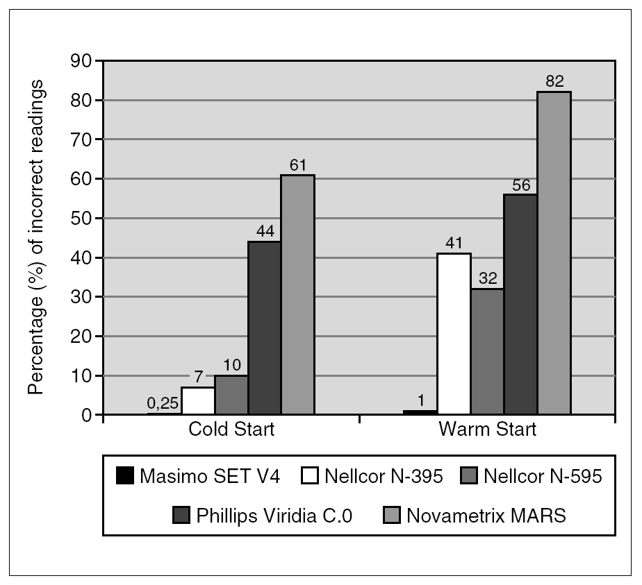
The adaptive probe of detection ("APOD") is also an "intelligent" algorithm, which helps decrease very significantly the percentage of incorrect readings as shown in figure 5. In the figure, it is shown that some monitors provide incorrect readings in about 60-80 % of the time, compared to the Masimo SET with "APOD", which only provides 0.25 % to a maximum of 1 % of incorrect readings with warm start, when the probe has been dislodged from the patient. The Masimo SET device can also be put into 'Max sensitivity' for times when perfusion is very low and clinicians at the bedside want to see what is going on and are not worried about the probe coming off the patient.

Figure 5. "APOD" is an "intelligent" detector, which also helps decrease very significantly the percentage of incorrect readings as shown.
Another innovation of Masimo is the "Signal IQ," which permanently verifies the quality of the signal, providing clinicians with a way to know when to question the displayed measurement. This is summarized in figure 6, where the first box shows the reading when there is absolutely no motion. Notice how the Signal IQ spike is high and displays the timing of each pulse. In Box A, there is some motion, but the Signal IQ is still very adequate, the bars are still high and pulse timing is indicated. In Box B with motion, Signal IQ is a little decreased, but the reading will continue to be accurate. Finally, in Box C with motion, Signal IQ spikes are almost absent. This alerts the clinicians to possibly corrupted SpO2 data, so that clinicians do not increase or titrate FIO2 on false premises.
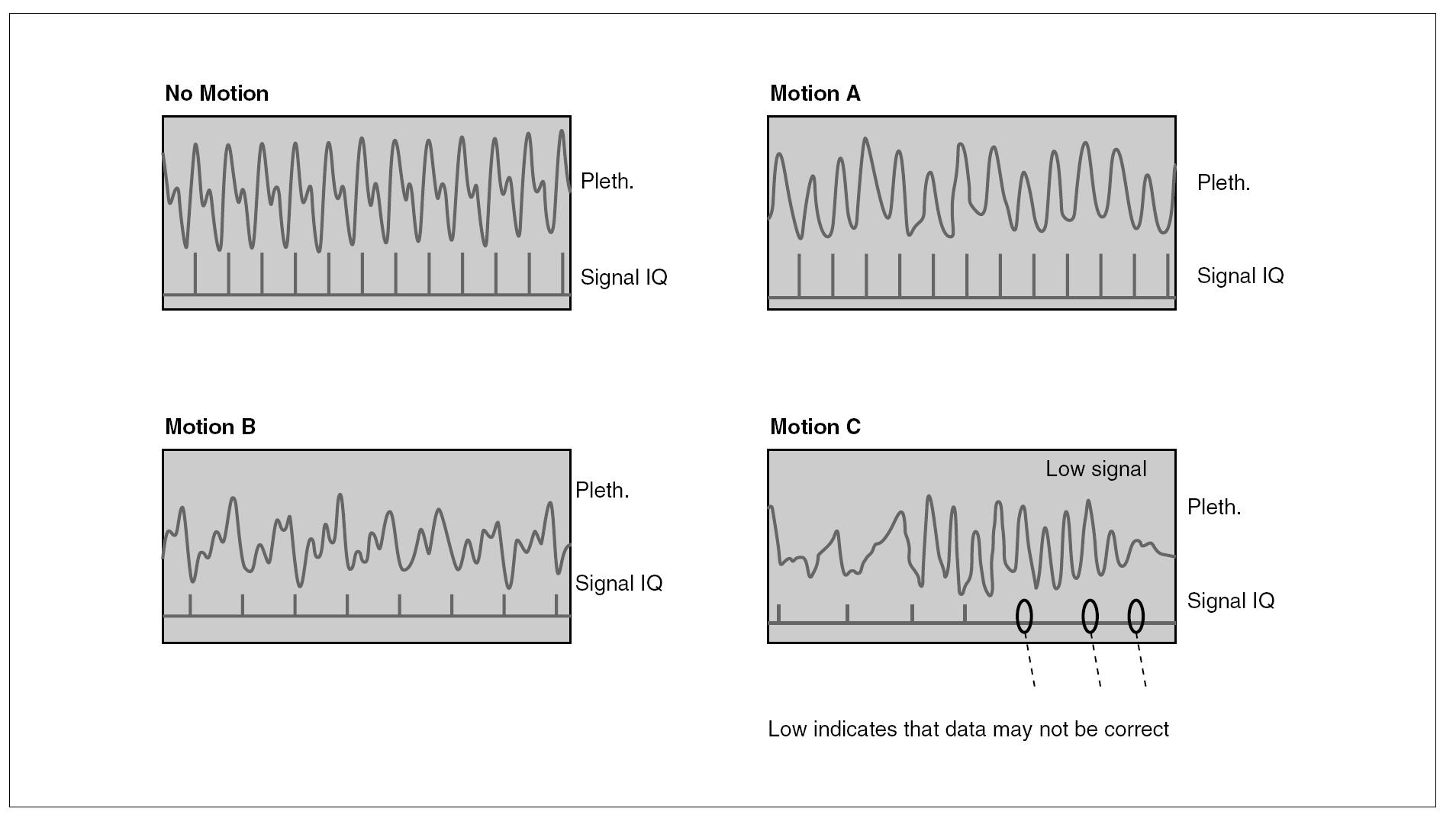
Figure 6. Signal IQ which permanently verifies the quality of the signal (see text).
Through innovative biophysics and technology, many other advances have been added to this new monitor. One of them is registered as "ClearVue," which essentially allows clinicians to "see well." Another one is the "SmartTone" that beeps in synchrony with the pulse, even during motion. Another one is "Diagnostic Pleth" which displays the true plethysmograph, while the other pulse oximeters distort the shape of the plethysmograph. Yet another significant advance is the "Perfusion Index". This one indicates the strength of the signal of the arterial pulse and could be of great value in diagnosing problems and severity of illness, even while oxygen saturation readings are accurately maintained.
Through all the advances described, Masimo SET SpO2 monitor has been proven to have the greatest sensitivity and specificity compared to any other pulse oximeter in the world, as shown later in comparative studies.
New sensor technology utilized in pulse oximetry
Another important invention of Kiani and others at Masimo Corporation are the sensors called low noise optical probes (LNOP). These are very useful, in addition to the adaptive filters, DST and SET described.
The LNOP sensors minimize the noises 1, 2, 3 and 4 mentioned above. They have a recessed photo detector in a pliable cavity that reduces motion noise, acting as a "shock absorber," reducing also the noise produced by ambient light, electro-surgical noise and the noise produced by the movement of the patient. These sensors are very light in terms of their minimal weight, in comparison with standard sensors and should reduce the weight put on the baby's foot or hand. In some cases, sequelae (i. e.: foot drop) have occurred at sensor site because of the weight of the conventional sensors and therapy was required to get the foot back to its normal position. The LNOP sensors are also very durable, and the adhesive has a very special characteristic that allows it to recover the adhesive properties if wiped with rubbing alcohol. Masimo has also recently introduced a new sensor, call LNOP Blue sensor, which is designed for cyanotic patients who have oxygen saturations in the range of 60 % to 80 %. Clinical studies are under way to see how much this new sensor improves SpO2 accuracy on these types of patients.
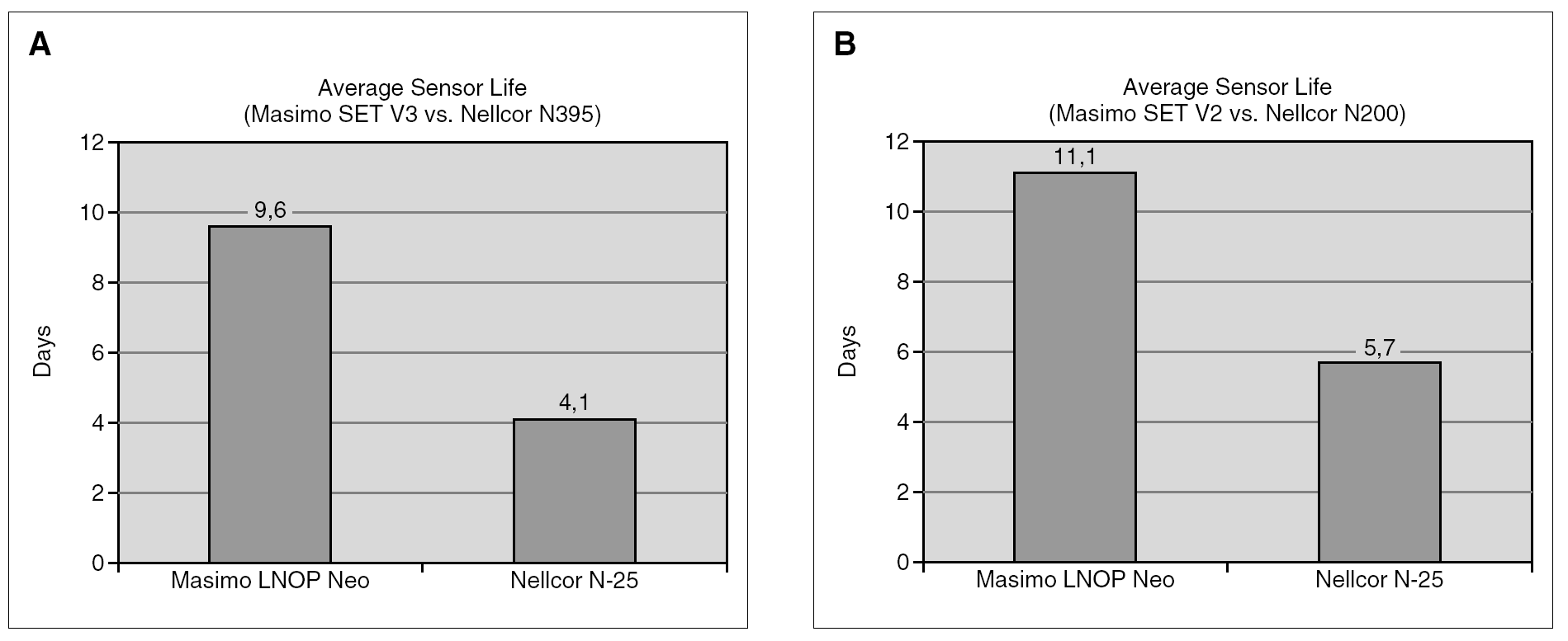
Figure 7 shows the average sensor life as reported by Wischniewski18 and by Thomas et al19. Masimo LNOP sensors average life was between about 10-11 days and for Nellcor sensors it was between 4 and approximately 6 days. Erler et al20 fairly recently reported that there was more that a two-fold increase in the life of Masimo versus Nellcor sensors, and that this difference was consistent between various care givers in multiple settings, with a significant potential for cost savings derived from the use of Masimo disposable sensors in neonatal routine care.

Figure 7. Average sensor life as reported by Wischniewski et al18 and Thomas et al19.
Summary of clinical trials evaluating performance of saturation monitors
Pulse oximetry manufacturers have introduced technologies that claim improved detection of hypoxemic events. In the last few years, many studies have compared monitor performance; some have already been mentioned in this manuscript. The literature clearly supports that when we are measuring saturation, one SpO2 monitor is not the same as another SpO2 monitor, and that the advances made in technological and scientific knowledge can not be ignored when caring for sick newborns. Because improvements in signal processing and data rejection algorithms do affect data reporting, clinicians do need to be aware of the real evidence, as reported in peer reviewed manuscripts, as opposed to claims made by manufacturers.
In 2002, Hay et al21 revealed that false alarms, missed events and data drop outs are markedly and significantly less with Masimo SET technology and were worse with Novametrix MARS. (fig. 8). Hay et al compared monitors in unsedated NICU infants who were on supplemental oxygen or mechanical ventilation. Compared with Nellcor, Masimo SET had 86 % fewer false alarms which were also shortened in duration, resulting in 92 % less total alarm time. False desaturations, data drop-outs, and false bradycardias were lowest for Masimo SET, as was the capture of true desaturations and bradycardias. Additionally, Masimo SET identified nearly all bradycardic episodes vs. 14 % for the Nellcor. Most notably, these so called "new-generation devices" differed greatly in their ability to detect changes in heart rate: the frequency of frozen pulse-rate during times of ECG heart rate changes was 0 (zero) only for Masimo21.
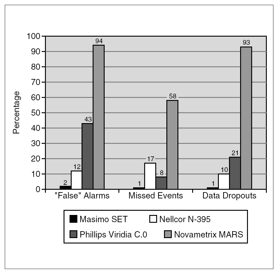
Figure 8. Hay et al compared monitors in unsedated NICU infants who were on supplemental oxygen or mechanical ventilation. The percentage of false alarms, missed events and data dropouts varied significantly.
Sahni et al22 compared Masimo versus Nellcor in different periods in infants undergoing pain due to circumcision. They found that there was significantly less variability and incidence of artifact with Masimo SET, and that the heart rate was more accurate with less procedural error. The amount of artifact was two to five times less, even 50 times less in some occasions, and there were significantly less false alarms. They concluded that monitoring with Masimo SET was continuous and much more precise in all the circumstances studied compared to Nellcor.
Goldstein et al23 reported that the monitors are also different in their ability to track significant heart rate changes. With Phillips HP, the heart rate changes were missed in 30 % of the occasions, with Nellcor N395 in 18 %, and with Masimo SET in only 1 %.
Barker24 reported on "motion-resistant" pulse oximetry and that the Masimo pulse oximeter demonstrated the best performance of 20 instruments tested.
Durbin and Rostow25 showed that Masimo SET was associated with lower failure rates, fewer arterial blood gas samples and shorter time in oxygen adult post surgery patients compared to the Ohmeda 374. This significant research was one of the first studies to compare the impact of different pulse oximetry technologies on patient processes and outcomes and also reported that Masimo SET reduced latent conditions, which are linked to medical errors25.
Robertson and Hoffman26 recently investigated the effects of signal integrity and saturation on data availability of three monitors. They compared data reporting and signal heuristic performance and agreement among the Nellcor N-395, Massimo SET and GE Solar 8000 Oximeters and the effects of conditions on signal integrity and arterial oxygen saturation. These were performed in a blinded side-by-side comparison of technologies. They found that with poor signals, with motion or during hypoxemia, the new oximeters are not clinically equivalent to each other or to the older Solar 8000 oximeter. They also found that agreement between the devices deteriorated in the presence of low signal quality, motion or hypoxemia. Masimo reported less data questionable than Nellcor in this blinded study26.
Kawagishia et al27, compared the failure times of pulse oximeters during cuff-induced hypo perfusion in volunteers. To evaluate the failure time of each pulse oximeter, time to the peak of blood pressure, time to loss of signal and time to recovery of signal and failure interval, the authors compared four monitors (Masimo SET, Nellcor N-395, Nellcor N-20PA and Nellcor D-25). The time to loss of signal was significantly longer in Massimo than any of the other pulse oximeters. Masimo also showed significantly shorter times to recovery of signal and the failure interval was markedly better and did not lose the signal as rapidly as the other oximeters studied. The observations in this study support that accurate data is more available with fewer false-positive alarms when using the Massimo SET oximeters27.
Bohnhorst et al28, performed a very revealing study in unsedated pre-term infants (median gestation age 25 weeks, with the range of 24-30 weeks) to detect the monitors reliability in detecting hypoxemia and bradycardia. For comparison, long-term recordings of TcPO2 and heart rate by other monitoring were used. The recordings were analyzed for episodes with TcPO2 less than 40 torr or with a heart rate less than 80 beats per minute for more than 5 seconds. In this study, there were a total of 202 falls in PO2; 5.4 % of the total hypoxemic episodes were missed by one the newer instruments (Nellcor) and only 0.5 % of the episodes were missed by the other (Masimo). Of interest was that of 54 total bradycardic episodes, only 14 were identified by all the oximeters studied; 69 % of the episodes were missed by the Nellcor monitor and only 7 % by the Masimo instrument. Thus, it appears that Nellcor's reduced false alarm rate is achieved at the expense of an unreliable and/or delayed identification of hypoxemia and bradycardia28.
In a recent investigation in an animal model of low perfusion caused by infection, Hummler29 had shown that low perfusion caused by emerging sepsis may result in inaccurate SpO2 measurements, but that the episodes in this animal model were less common with the Masimo SET vs. the Nellcor Oxismart XL. They showed that there were fewer episodes with a false SpO2 reading using the Masimo SET as verified by cooximetry and the average bias (SpO2-SaO2) was also significantly different between the two devices.
The new signal extraction technology has also been compared by Irita et al30 during hypothermic cardiopulmonary bypass with non-pulsatile flow. The Masimo SET displays accurate SpO2 significantly more frequently and longer than a conventional oximeter, showing its usefulness for monitoring SpO2 during hypoperfusion30. In addition, "Perfusion Index" indicates the strength of the signal of the arterial pulse and could be of great value in diagnosing problems and severity of illness31.
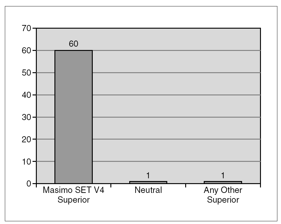
These and other authors have also done several comparative studies. Through independent and objective literature summary, we summarize in figure 9, the number of studies to date comparing Masimo SET versus any other oximeter. As it can be seen, Masimo has been proven superior in all cases but two; in one study the evaluation was neutral, not showing any benefit of the other monitor. The immense majority of independent and objective studies did not find any monitor to be superior to Masimo SET. (We provide a list of pertinent bibliography for anyone interested in extensive detailed review.)

Figure 9. Comparison of available studies.
Concluding comments
The technology of pulse oximetry has been shown to be effective in helping clinicians detect hypoxemia but we need to be more aware regarding hyperoxemia. Many monitors have difficulty obtaining accurate readings during periods of motion, low perfusion and "noise" resulting in frequent nuisance alarms or false readings. The so called "next-generation" technologies of SpO2 monitors are marketed as being able to obtain accurate values when former conventional technologies cannot. The questions we should ask ourselves as clinicians are whether these technologies live up to the supplier's claim. As clinicians, we need to know the ability of each monitor to precisely reduce nuisance alarms, evaluate the evidence of missed alarm events and many other factors that have been discussed in this manuscript. It is only following objective, peer reviewed evidence that we as clinicians should (a) understand the differences in different monitors and (b) offer to our patients the most precise technology for their own well being. The evidence available in peer reviewed manuscripts should take precedence when health care providers and facilities make implementation decisions for the care of their patients. Additionally, we should be aware of the evidence about the lower cost of the best technology. Several hospitals in the USA claim to save > 100,000 dollars or more per year after changing to Masimo SET technology. SpO2 monitors with greater and refined accuracy to resolve most artifacts and to increases sensitivity at low blood flow, pulse amplitude and blood oxygenation values, ensure better clinical care, decrease the need of measuring PaO2 and hopefully, one day, may make it obsolete.
Based on the available scientific evidence we recommend the use of reliable, new SET SpO2 monitoring, avoid responding to "false alarms" and keep alarm limits as long as the infant is receiving FiO2 > 0.21 during high risk periods of development, starting in the delivery room from the time of birth. Finally, we also recommend managing the infant with O2Sat which, by known physiologic relations between hemoglobin and oxygen, will lead to PaO2 that is "not low nor high" by currently accepted standards.
Utilization of the most adequate SpO2monitoring in the NICU can improve clinician confidence in SpO2 values, leading to a more judicious dosing of oxygen with possible reductions in hypoxic and hyperoxic side effects that have been unfortunately common in neonatology. The most trustworthy technology also leads to fewer confirmatory arterial blood gas analyses, faster weaning from mechanical ventilation and lower costs.
We know that no one knows what the "best" or "ideal" saturation range is for all preterm infants of all gestational ages, at all postnatal ages. This needs detail study. However, eradicating some bad practices is not the same as implementing routinely unproven practices in a rigid way. Increase awareness of known and proven facts will decrease the gap between knowledge and practice. Paying attention to "details" we can accomplish a clinical difference and improve outcomes.
Acknowledgements
We are thankful to Mr. J. Kiani for the critical review of the manuscript and helpful suggestions on technical aspects and to the Goddard Scholarship at Emory Children's Research Center for the support to one of the authors (A.S)
Dr A. Sola lectures in relation to oxygen therapy, oxygenation levels and pulse oximetry. Masimo Corporation provides funds for his travel and honorarium 4-8 times per year and is also one of several sponsors in anual educational post-graduate courses organized by Dr. Sola.

