
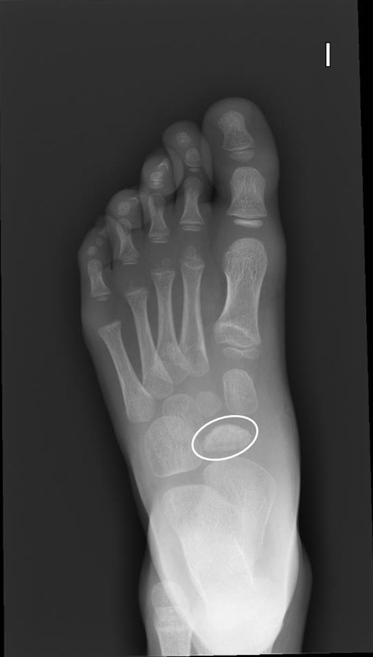
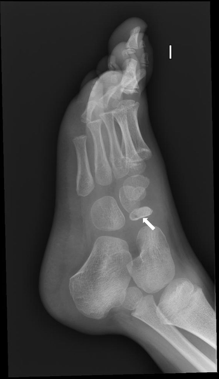
A boy aged 4 years presented with a left-sided limp of several weeks’ duration. The episodes of limping were intermittent, occurring predominantly in the morning, and did not limit his daily activities. The patient denied any trauma, overexertion, or recent infection. The physical examination was unremarkable, except for tenderness on palpation in the medial midfoot region, with no associated signs of inflammation or restriction of joint mobility. Plain radiographs of the left foot (Figs. 1 and 2) ruled out fracture. However, the images revealed cortical irregularity, increased density (white arrow), and wafer-like flattening of the tarsal navicular bone (white circle), findings consistent with Köhler disease.


Köhler disease is an osteochondrosis caused by avascular necrosis of the proximal ossification center of the navicular bone. It is more frequent in boys (5:1), and its incidence peaks between ages 3 and 7 years.1,2 Although its etiology has not been fully elucidated, it is associated with insufficient blood flow, as the navicular bone is the last tarsal bone to ossify. It is a self-limiting disease. The management is conservative and focuses on pain relief and relative rest, although some cases may require immobilization with a cast to reduce the mechanical load. Early diagnosis and adequate follow-up are required to detect potential complications.3