


Histological studies in people with cystic fibrosis (pwCF) show appendiceal mucosal hyperplasia and thick secretions, which cause dilatation, usually in absence of significant inflammation.1 Ultrasound (US) reveals an abnormal appendiceal diameter (AD >6 mm) in 20%–80% of asymptomatic pwCF.2 Although the incidence of appendicitis is lower in this group compared to the general population (1%–2% vs 7%), its diagnosis is often delayed, increasing the risk of perforation or abscess.1–3 As cystic fibrosis transmembrane conductance regulator (CFTR) is expressed in intestinal epithelial cells and ganglia,4 CFTR modulators, and the elexacaftor/tezacaftor/ivacaftor (ETI) combination in particular, may reduce the AD.5 We evaluated the impact of ETI on the AD and its association with changes in the sweat chloride concentration.
We conducted a single-center ambispective longitudinal observational study (Ethics Committee file No. 2022.102) that adhered to the principles of the Declaration of Helsinki principles. The inclusion criteria were having a diagnosis of CF, current treatment with ETI, and a baseline AD of 6 mm or greater on abdominal ultrasound or magnetic resonance enterography (MRE). The exclusion criteria were having any contraindication to CFTR modulators, a previous history of appendectomy, a normal baseline AD, or lack of pretreatment imaging (supplemental material, Figure S1). Baseline imaging reflected the routine clinical surveillance for hepatobiliary/pancreatic involvement, which systematically includes intestinal/appendiceal evaluation when feasible; an MRE was performed in one patient due to the presence of gastrointestinal symptoms and elevated fecal calprotectin. Ultrasound was performed as part of routine practice using a Canon Aplio 550 system with linear transducers (3−8 MHz) by one of three pediatric radiologists (with 7–17 years of experience); images were not systematically double-read. In addition, as these were surveillance studies in asymptomatic patients, there were no indirect sonographic signs of appendicitis (such as periappendiceal hyperechoic fat, appendiceal wall hyperemia, or free fluid). Sweat chloride was measured by the Gibson-Cooke method (standardized pilocarpine iontophoresis and quantitative analysis), in the morning (09:00−12:00) and before administration of the ETI dose during the pretreatment visit, while patients were clinically stable on their usual maintenance therapies. Data were managed through the REDCap platform. We have expressed quantitative data as median and interquartile range (IQR). Normality was assessed using the Shapiro-Wilk test and kurtosis tests. We compared pre- and post-treatment data for variables that met the normality assumption using the t test for paired samples with the STATA software, version 18.0. Statistical significance was set at P < .05.
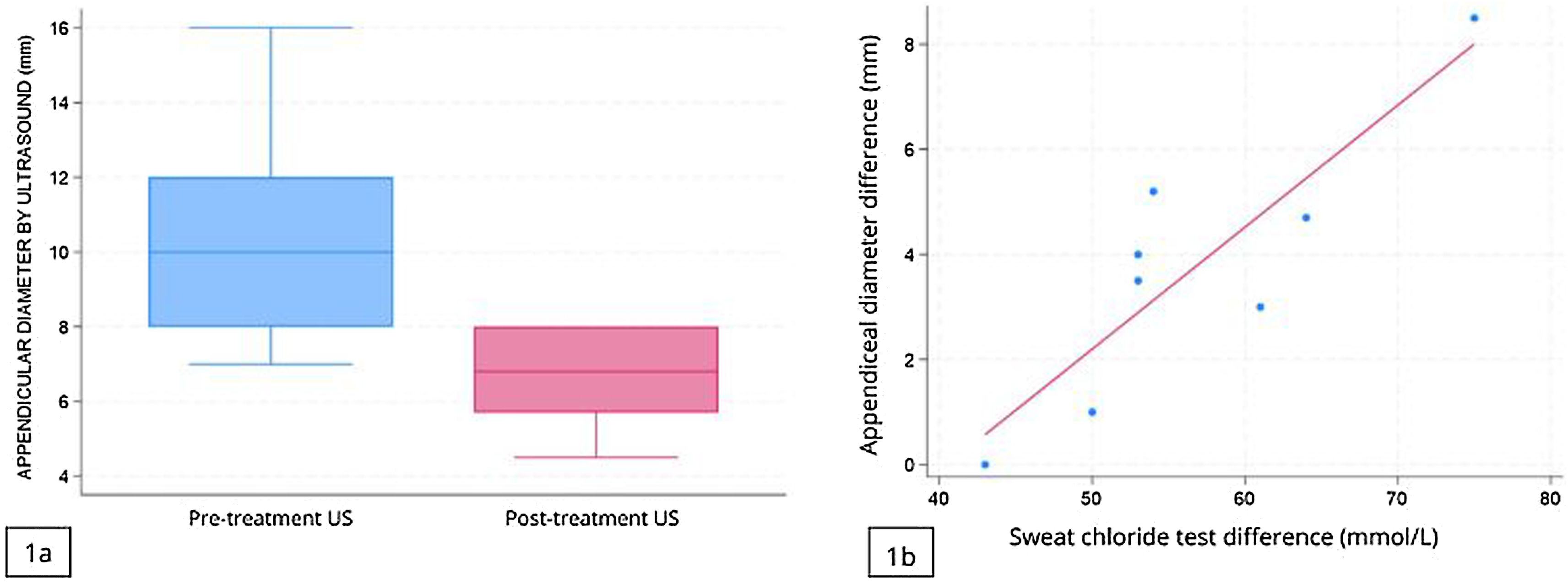
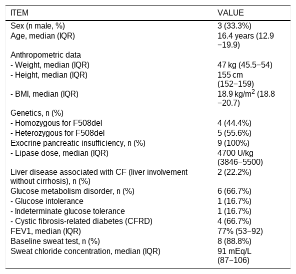
Nine patients were recruited (66.7% female), aged 8.5–24.9 years, all with pancreatic insufficiency (fecal elastase <200 µg/g). Four (44.4%) had prior exposure to CFTR modulators (one to lumacaftor/ivacaftor; one to lumacaftor/ivacaftor then tezacaftor/ivacaftor; two to tezacaftor/ivacaftor). Table 1 summarizes additional characteristics of the sample. The baseline imaging (ultrasound in 8 patients, MRE in 1) was performed a median of 0.9 years (IQR, 0.5–3.7) before ETI initiation, with a mean AD of 10.4 mm (SD, 2.7). Follow-up ultrasound scans were performed between 1.1 and 2.7 years (median, 2.5; IQR, 2.1–2.6) post-ETI. The AD decreased by a mean of 3.8 mm (95% CI, 1.9–5.7; P = .0017), reaching normal values (≤6 mm) in 44.4% of the patients (Fig. 1a). Patients with persistent abnormal ADs were older (median age, 19.9 [IQR 15.5–21.8] vs 14.6 [IQR, 12.4–16.4] years) and had larger baseline ADs (mean, 11.8 [SD, 2.9] vs 8.8 [SD, 1.5] mm), although these differences were not significant (P > .05) (Supplemental material, Figure S2).
Description of the study sample.
| ITEM | VALUE |
|---|---|
| Sex (n male, %) | 3 (33.3%) |
| Age, median (IQR) | 16.4 years (12.9−19.9) |
| Anthropometric data | |
| - Weight, median (IQR) | 47 kg (45.5−54) |
| - Height, median (IQR) | 155 cm (152−159) |
| - BMI, median (IQR) | 18.9 kg/m2 (18.8−20.7) |
| Genetics, n (%) | |
| - Homozygous for F508del | 4 (44.4%) |
| - Heterozygous for F508del | 5 (55.6%) |
| Exocrine pancreatic insufficiency, n (%) | 9 (100%) |
| - Lipase dose, median (IQR) | 4700 U/kg (3846−5500) |
| Liver disease associated with CF (liver involvement without cirrhosis), n (%) | 2 (22.2%) |
| Glucose metabolism disorder, n (%) | 6 (66.7%) |
| - Glucose intolerance | 1 (16.7%) |
| - Indeterminate glucose tolerance | 1 (16.7%) |
| - Cystic fibrosis-related diabetes (CFRD) | 4 (66.7%) |
| FEV1, median (IQR) | 77% (53−92) |
| Baseline sweat test, n (%) | 8 (88.8%) |
| Sweat chloride concentration, median (IQR) | 91 mEq/L (87−106) |
Abbreviations: BMI, body mass index; CF, cystic fibrosis; FEV1, forced expiratory volume at one second; F508del, delta F508 mutation.
We observed a strong correlation between sweat chloride concentrations and changes in AD (Pearson r, 0.87; P = .0048) (Fig. 1b); for every unit decrease in sweat chloride, the AD decreased by 0.2 mm (b = 0.2; P = .005; 95% CI, 0.1−0.3). Fecal calprotectin decreased significantly from a baseline median of 95 µg/g (IQR, 56–170; range, 16–459) to a follow-up median of 31 µg/g (IQR, 11–55) (P = .0117). Baseline calprotectin levels were elevated (>50 µg/g) in 77% of patients (7/9), with the percentage decreasing to 33% (3/9) at follow-up, a difference that was not statistically significant (P = .1250).
Our study suggests that AD decreases significantly after ETI initiation, with nearly half of patients achieving normalization. The correlation between the reductions in sweat chloride concentration and in AD suggests that improved CFTR functionality directly reduces intestinal secretions and appendiceal size, which supports the role of sweat chloride concentration as a biomarker of systemic and extrapulmonary therapeutic responses.4,6 At the same time, the significant decrease in fecal calprotectin indicates a reduction in intestinal inflammation, which might affect appendiceal size, although the sample size precluded drawing firm conclusions.
While AD alone is not sufficient to diagnose appendicitis, baseline monitoring facilitates abdominal pain assessment, particularly for clinicians unfamiliar with cystic fibrosis presentations. As highly effective CFTR modulators reduce AD, the risk of appendicitis and its clinical presentation in pwCF may shift to resemble those of the general population, challenging historical assumptions of a lower incidence.1
The limitations of the study include the small sample size and selection bias due to the exclusive inclusion of patients with abnormal baseline diameters, which restricts extrapolation. Baseline imaging relied on routine surveillance practices rather than symptom-driven protocols (except for one MRE). Furthermore, ultrasound is operator-dependent; and since this real-world study was ambispective, we did not evaluate interobserver variability.
To our knowledge, this is the first study to demonstrate a reduction in AD following treatment with ETI and its association with improvements in the sweat test. In conclusion, ETI therapy reduces AD in pwCF, achieving its normalization in nearly half of the cases. Furthermore, the magnitude of appendiceal size reduction is directly correlated to improvements in sweat chloride levels.
Informed consentThe data used in this study were collected and stored in an anonymized database. All participants aged 12 and 18 years assented to participation in the study, and adult patients and parents of minors provided signed informed consent to participation.
Ethical considerationsThe study was approved by the Ethics Committee of the Principality of Asturias under protocol number 2022.102.
FundingThe study did not receive any external funding.
The authors declare no conflicts of interest.
The following are Supplementary data to this article: