A girl aged 7 years was referred for assessment of chronic diarrhea and intermittent episodes of rectal bleeding with onset a year prior. The initial evaluation revealed mild elevation of fecal calprotectin (maximum, 118.2μg/g), so the evaluation was extended with an upper gastrointestinal endoscopy and an ileocolonoscopy.
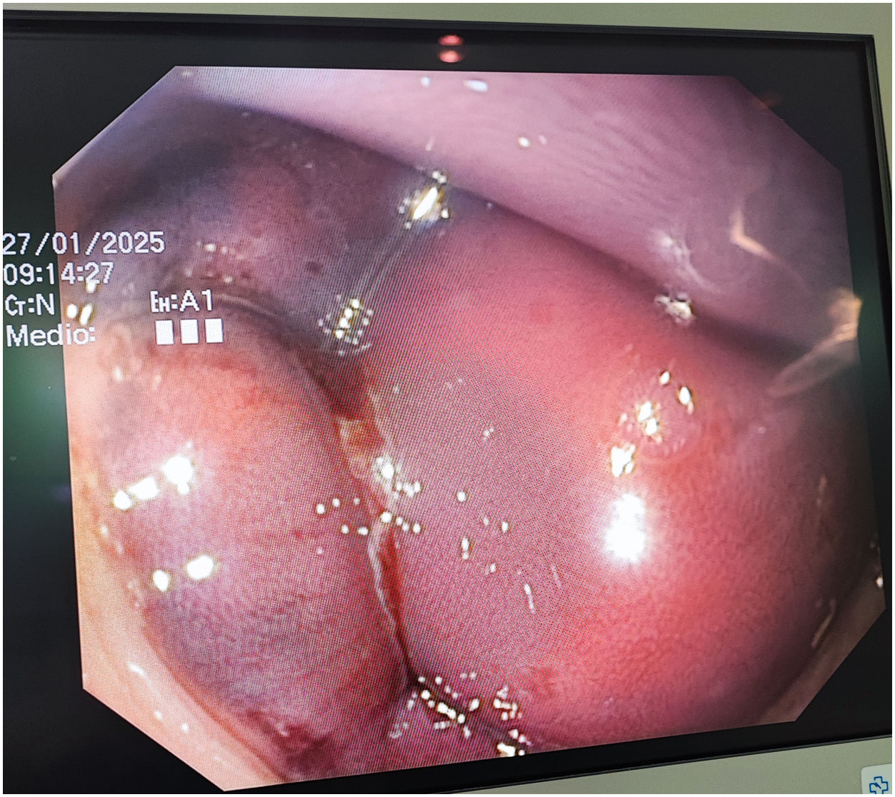
Both procedures were performed without incident under sedation and analgesia with propofol and fentanyl. As the colonoscope was withdrawn, several purplish submucosal lesions were visualized in the descending colon that had not been present during its initial advancement (Fig. 1).1,2 The patient remained stable, and laboratory tests showed no evidence of anemia or coagulation abnormalities.

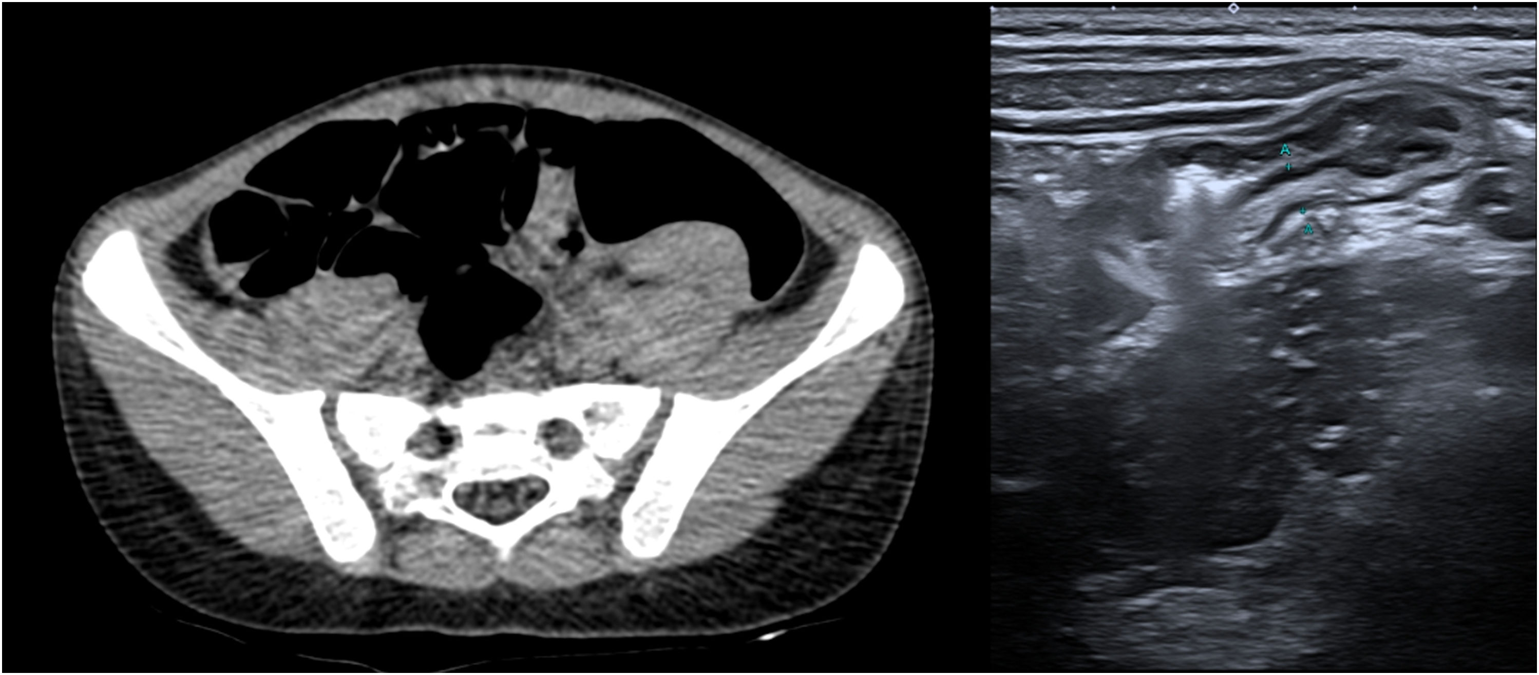
Computed tomography and ultrasound scans of the abdomen revealed multiple nodular lesions attached to the colonic wall (Fig. 2).3 At 48h, the patient had an isolated and self-limited episode of rectal bleeding. Due to the suspicion of iatrogenic submucosal colonic hematomas, the care team opted for a conservative approach,1,2 with subsequent evidence of progressive resolution.

Colonoscopy is a key diagnostic tool that is widely used in pediatric care, and it has a high safety profile, although it is not free of complications. Colonic submucosal hematomas are rare and usually associated with trauma during the examination, although they can develop even in the absence of obvious manipulation. Their endoscopic detection helps avoid unnecessary interventions.