

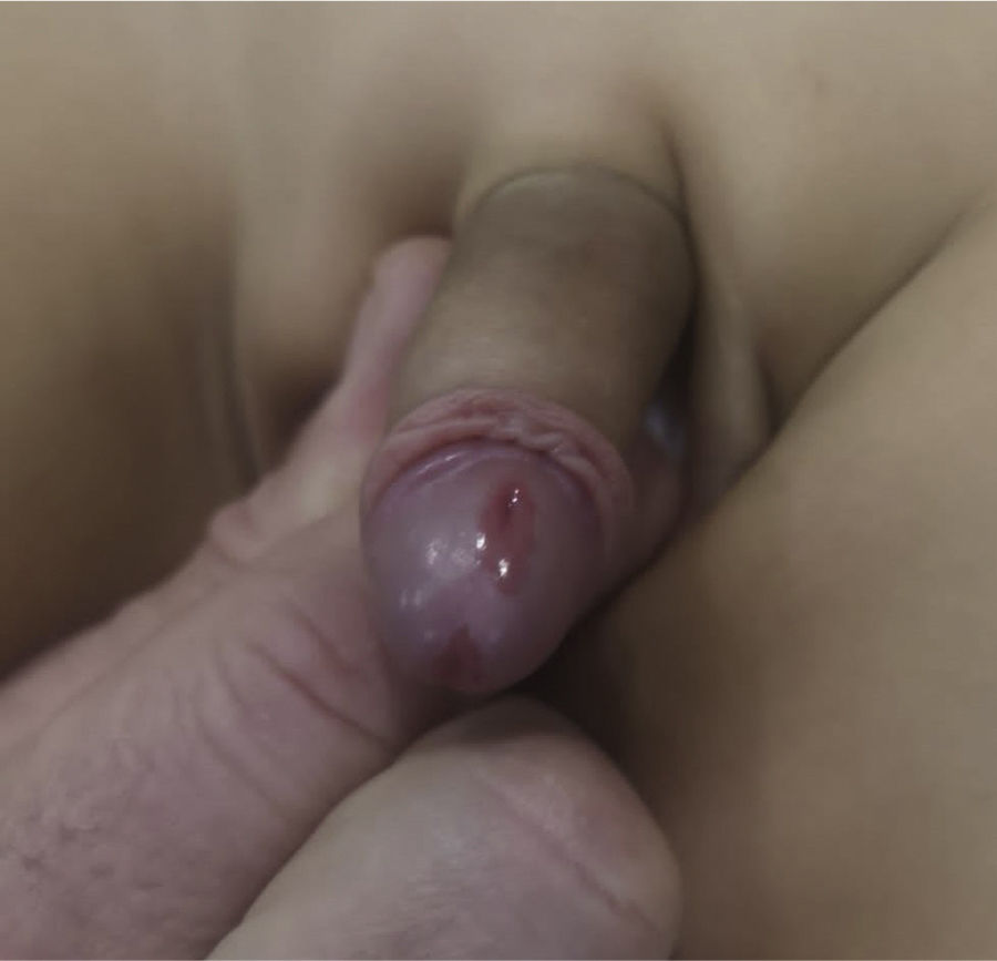
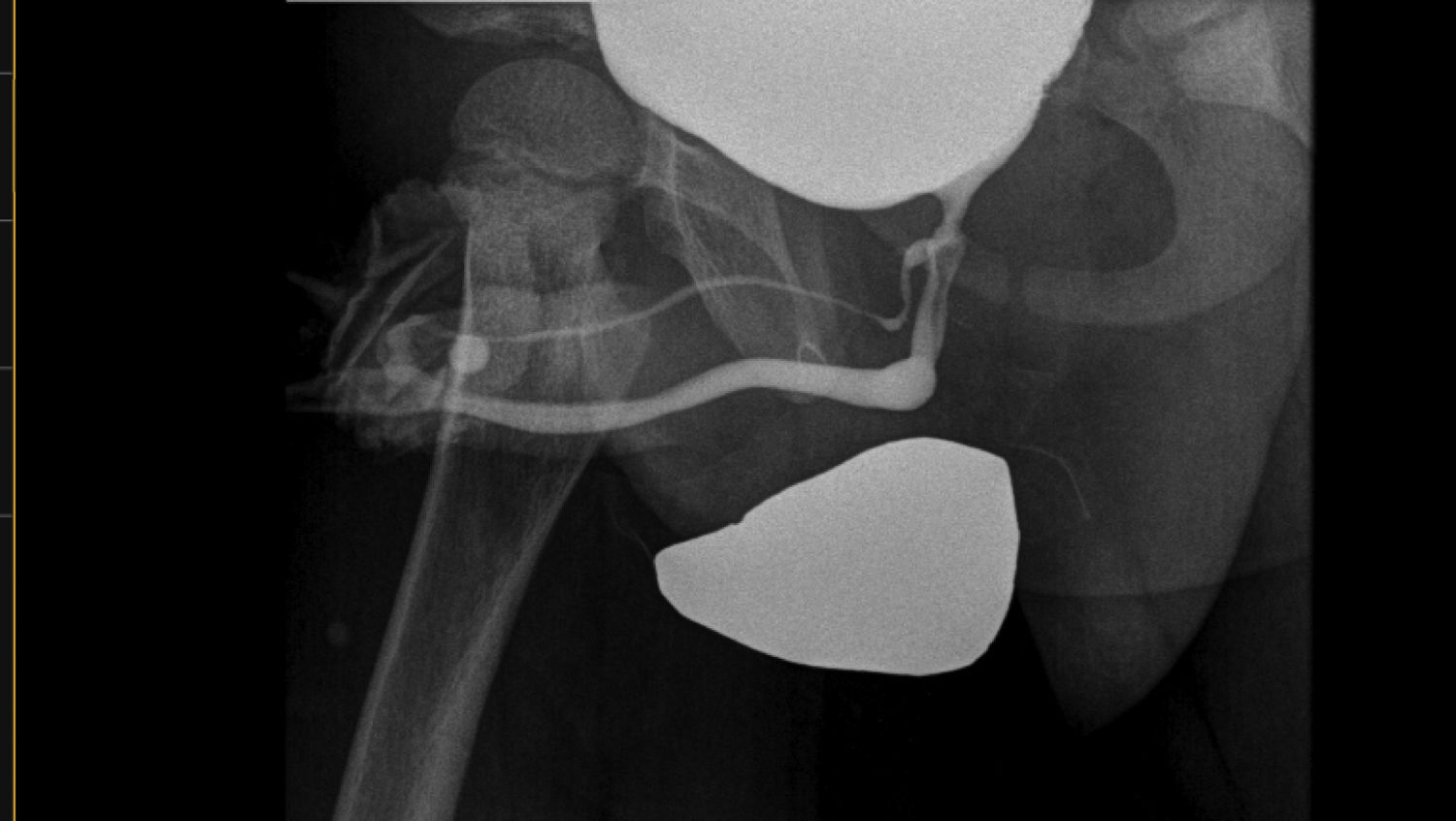
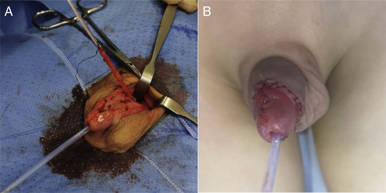
We present the case of a boy aged 5 years referred to the paediatric urology clinic for assessment of a double urethral meatus, which was asymptomatic: there was no passage or dribbling of urine through the dorsal meatus. Two meatuses were observed in the physical examination, one in the normal anatomical location and in a dorsal location (Fig. 1). The voiding cystourethrogram confirmed complete urethral duplication with a normal urethra and an accessory urethra originating from the main prostatic urethra at the level of the verumontanum (Fig. 2), corresponding to type 2 coronal complete urethral duplication in the classification of Lima et al.1 The patient underwent surgical intervention with circumcision, dissection and excision of the dorsal duplicated urethra, measuring 5cm in length, with ligation of the base (Fig. 3). An 8 Fr urinary catheter was also inserted. The patient had a favourable outcome in the immediate postoperative period, allowing removal of the urinary catheter, and was discharged 24h after the intervention. In the follow-up visits, it was reported that he remained asymptomatic.
 meatus and an accessory (dorsal) meatus.")

Voiding cystourethrogram: type 2 complete coronal urethral duplication, according to the classification of Lima et al.1
 Surgical intervention: dissection and removal of duplicated urethra. (B) Postoperative outcome.")
Urethral duplication is a rare congenital anomaly of unknown incidence. There are different classifications. The earliest ones were proposed by Williams and Kenawi2 in 1975 and by Effmann et al.3 in 1976 and, more recently, in 2016, Lima et al.1 proposed an anatomical-functional classification. It is a clinical spectrum that ranges from the absence of symptoms to the development of infection, urinary incontinence and penile curvature.
A physical examination of the genitals and perineum is necessary to suspect the diagnosis, with visualization of a second meatus, noting its location and likely trajectory.
The anatomy of the duplicated urethra can be determined by means of imaging tests with contrast and cystourethrography, allowing differentiation of the main urethra and the accessory one and assessment of the integrity of the former for the purposes of planning the surgical management.

