Journal Information
Vol. 99. Issue 5.
Pages 362-363 (01 November 2023)
Vol. 99. Issue 5.
Pages 362-363 (01 November 2023)
Images in Paediatrics
Full text access
Self-healing juvenile cutaneous mucinosis
Mucinosis cutánea juvenil autorresolutiva
Visits
707
Elvira Reneses Prieto
Corresponding author
ereneses@ucm.es

Corresponding author.
, Agustín López López, Elena M.ª López-Negrete Arenal, Inmaculada Olmedo Díaz
Hospital Puerta de Hierro Majadahonda, Madrid, Spain
This item has received
Article information
Full Text
Bibliography
Download PDF
Statistics
Figures (3)
Show moreShow less
Full Text

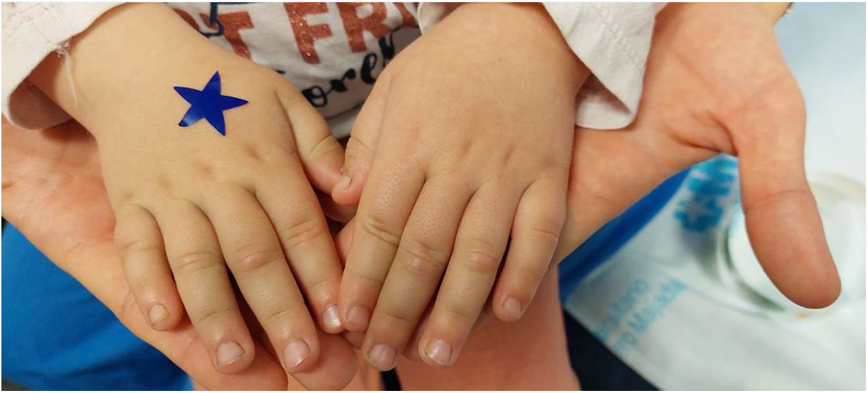
A girl aged 3 years presented with palmar pain of a few days’ duration that did not respond to commonly used analgesics. She was admitted for evaluation, with malignant disease ruled out first. In the first days of the stay, she developed hypertension, which responded to amlodipine. At 1 week from admission, the intense pain persisted, accompanied by inflammation, rigidity and loss of function in several joints, more severe at the interphalangeal level. Steroid therapy was initiated, after which the patient developed cutaneous nodules in different locations, as can be seen in Figs. 1 and 2.

Figure 1.

Frontal nodular lesions.

(0.11MB).
Figure 2.

Interphalangeal nodular lesions.

(0.09MB).

Several diagnoses were possible, so a biopsy sample was obtained from one of the lesions, the examination of which revealed mucin deposition in the dermis (Fig. 3), fibroblast and fibrovascular proliferation and lymphocytic inflammation, leading to the definitive diagnosis of self-healing juvenile cutaneous mucinosis, a rare childhood disease presenting with cutaneous and rheumatic manifestations. The cutaneous manifestations may include nodules in periarticular regions and the head or neck and the papules described as whitish lesions in erythematous cutaneous tissue.1

Figure 3.

Biopsy sample of a cutaneous lesion in the patient. Alcian blue stain evincing mucin deposition.

(0.14MB).

Despite its benign course, the abrupt onset of symptoms and the extensive cutaneous involvement may lead to the performance of an excessive number of diagnostic tests and overtreatment, at times aggressive, before the definitive diagnosis is reached.2,3

References
[1]
A. Bishnoi, A.K. Jindal, G. Anjani, P.K. Patra, D. Chatterjee, K. Vinay, et al.
Self-healing juvenile cutaneous mucinosis, a sclerodermoid disorder simulating juvenile dermatomyositis: a case-based review.
Rheumatol Int, 40 (2020), pp. 1911-1920
[2]
E.W. Cowen, G.A. Scott, M.G. Mercurio.
Self-healing juvenile cutaneous mucinosis.
J Am Acad Dermatol, 50 (2004), pp. 97-100
[3]
I. Luchsinger, J. Coulombe, F. Rongioletti, M. Haspeslagh, A. Dompmartin, I. Melki, et al.
Self-healing juvenile cutaneous mucinosis: clinical and histopathologic findings of 9 patients: the relevance of long-term follow-up.
J Am Acad Dermatol, 78 (2018), pp. 1164-1170
Copyright © 2023. Asociación Española de Pediatría
Idiomas
Anales de Pediatría (English Edition)
Article options
Tools
es en

¿Es usted profesional sanitario apto para prescribir o dispensar medicamentos?

Are you a health professional able to prescribe or dispense drugs?