

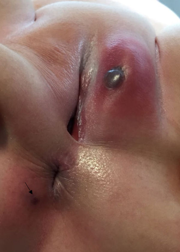
A previously healthy girl aged 15 months with no family history of interest who attended a childcare facility received a diagnosis of lobar pneumonia that responded favourably to treatment with amoxicillin at a dose of 90mg/kg/day. On day 10 of treatment, the fever recurred. A hard mass with an erythematous base and a necrotic central area with a bullous lesion was detected in the genitals (Fig. 1). There was also a small, purplish necrotic lesion in the anal margin. Both lesions worsened over the next 24hours. The laboratory tests revealed severe neutropenia (420/μL) and elevation of liver enzymes (AST/ALT, 43/102 U/L) and C-reactive protein (244.6mg/L). This led to diagnosis of ecthyma gangrenosum and initiation of antibiotherapy with ceftazidime, amikacin and vancomycin. Pseudomonas aeruginosa was isolated from culture of a sample of the cutaneous lesion, and there was no growth in the blood culture. Treatment with ceftazidime and amikacin continued for a total of 21 days. The response was favourable, with full resolution of the genital lesion and re-epithelialization of the necrotic area. The neutrophil count normalised, and there has been no evidence of immunodeficiency to date (normal respiratory burst and immunophenotyping of peripheral lymphocytes).
.")
Ecthyma gangrenosum is an infrequent infectious disease manifesting with a papular or nodular lesion that progresses rapidly to necrotic ulceration with a black central crust.1,2 It is typically associated with infection by P. aeruginosa, although other aetiological agents may be involved, chiefly Staphylococcus aureus.2,3 Early detection and initiation of antibiotherapy are key prognostic factors.1 This disease is frequently associated with neutropenia and immunodeficiencies, so their presence must always be ruled out.1,3

