

In April 2020, a new systemic inflammatory syndrome was first described in the paediatric population with features that overlapped those of Kawasaki disease, toxic shock syndrome and macrophage activation syndrome.1 An association with SARS-CoV-2 was rapidly established, as most affected patients tested positive for the virus and/or had clinical manifestations compatible with COVID-19 2–6 weeks prior to onset.
One of the most frequent features of the syndrome is the presence of gastrointestinal symptoms (abdominal pain, vomiting and diarrhoea), in some cases compatible with acute abdomen. This suggested the possibility of an association between the multisystemic inflammatory syndrome in children (MIS-C), SARS-CoV-2 infection and acute appendicitis.2–4
We present the cases of 3 children with an unremarkable prior history that developed acute appendicitis in the context of MIS-C. Table 1 summarises the clinical and laboratory characteristics of these cases.
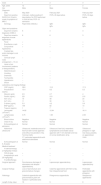
Clinical characteristics, laboratory values, imaging features and treatment of patients with MIS-C (criteria of the Centers of Disease Control and Prevention) and acute appendicitis.
| Case | 1 | 2 | 3 |
|---|---|---|---|
| Age, years | 12 | 2 | 2 |
| Sex | Male | Male | Female |
| Diagnosis | |||
| Date | May 2020 | February 2021 | February 2021 |
| Days from positive SARS-CoV-2 test to onset of symptoms of MIS-C | Unknown, mother positive 21 days before. No PCR performed in child at time, PCR– at admission | PCR+ 26 days before | PCR+ 33 days before |
| Serology | Rapid total antibody + | IgM+ | IgM+ |
| IgG+ | IgG+ | ||
| Signs and symptoms | |||
| Days from onset to diagnosis of MIS-C | 6 | 3 | 3 |
| Days from onset to diagnosis of acute appendicitis | 8 | 6 | 3 |
| Fever | + | + | + |
| Exanthema o rash | + | + | + |
| Conjunctival hyperaemia | + | + | + |
| Cracked lips and/or damage to oral mucosa | – | + | + |
| Cervical lymph node enlargement > 1.5 cm | – | – | – |
| Hand or foot involvement (oedema or desquamation) | – | – | – |
| Abdominal pain | + | + | + |
| Vomiting | + | – | – |
| Diarrhoea | + | + | – |
| Tachycardia | – | + | + |
| Hypotension | – | – | – |
| Shock | – | – | – |
| Laboratory and imaging findings | |||
| CRP (mg/dL) | 38.8 | 13.5 | 17.3 |
| Procalcitonin (ng/mL) | 10.4 | 8.2 | 0.2 |
| Albumin (g/dL) | 2.9 | 2.7 | 4 |
| Ferritin (ng/mL) | 221 | 213 | 210 |
| AST (U/L) | 19 | 48 | 23 |
| ALT (U/L) | 11 | 30 | 14 |
| D-dimer (μg/mL) | 5.3 | 4.2 | 3.9 |
| BNP (pg/mL) | 159 | 82 | 14 |
| Troponin (pg/mL) | 1 | 7 | 1 |
| Leucocytes (103/mm3) | 14.82 | 12.94 | 18.11 |
| Lymphocytes (103/mm3) | 0.75 | 1.69 | 2.09 |
| Blood culture | Negative | Negative | Negative |
| Chest X-ray | Normal | Normal | Normal |
| Echocardiogram at admission | Normal | Normal | Normal |
| Abdominal ultrasound/ | Ultrasound: moderate amount of free fluid with normal appendix and without visualization of tip. Terminal ileitis | Ultrasound: free fluid, mesenteric lymphadenitis and dilated caecal appendix with 7 mm diameter and loss of normal stratification at tip. | Ultrasound: phlegmon in right iliac fossa, possibly an appendiceal mass. |
| CT: perforated appendicitis with abdominal abscesses | |||
| Echocardiogram at 6−8 weeks | Normal | Normal | Normal |
| Medical treatment | |||
| IV steroid therapy (2 mg/kg/day 5 days) | – | + | – |
| IVIG (2 g/kg) | + | + | – |
| Acetylsalicylic acid (50 mg/kg/day) | – | + | – |
| IV antibiotherapy | + | + | + |
| Surgical approach | Percutaneous drainage of abscesses and delayed appendectomy. | Laparoscopic appendectomy | Laparoscopic appendectomy |
| Surgical findings | Loss of wall structure and granulomatous changes | Catarrhal appendicitis with fibrin at tip, free intraperitoneal fluid | Perforated appendicitis with phlegmon |
| Pathology | Catarrhal appendicitis with granulomatous giant cell reaction in perforated area | Phlegmonous appendicitis | Gangrenous appendicitis |
| Length of stay, days | 18 | 9 | 8 |
ALT, alanine aminotransferase; AST, aspartate aminotransferase; BNP, brain natriuretic peptide; CPR, C-reactive protein; CT, computed tomography; IVIG, intravenous immunoglobulin; MIS-C, multisystemic inflammatory syndrome in children; PCR, polymerase chain reaction.
Case 1. Boy aged 12 years that presented to the emergency department in May 2020 with a fever of 3 days’ duration, abdominal pain, vomiting and diarrhoea associated with a transient, salmon-pink rash. The workup revealed marked elevation of acute phase reactants (APRs) and the SARS-CoV-2 polymerase chain reaction (PCR) test was negative. The initial ultrasound revealed terminal ileitis with a normal appendix and a small amount of free intraperitoneal fluid. The patient started antibiotic treatment, and the fever persisted. Systemic inflammatory syndrome in children was suspected due to the history of recent infection in the mother and a positive rapid SARS-CoV-2 total antibody test. Intravenous immunoglobulin (IVIG) was administered, which achieved resolution of the fever but with worsening of the abdominal pain associated with features suggestive of acute abdomen. The patient underwent an abdominal computed tomography (CT) scan, which revealed perforated appendicitis and several abdominal abscesses. The absences were drained percutaneously, followed by gradual improvement. The patient underwent a delayed appendectomy at 3 months of follow-up without complications.
Case 2. Boy aged 2 years that presented with fever of 48 h’ duration, malaise, abdominal pain, diarrhoea, exanthema and cracked lips (Fig. 1A). The patient had a history of oligosymptomatic COVID-19 4 weeks before. Elevation of APRs, including ferritin and D-dimer levels. The clinical and laboratory features were indicative of MIS-C, leading to initiation of steroid therapy and IVIG. The patient was afebrile for 24 h, which subsequent recurrence of fever and worsening of abdominal pain, which was located in the right iliac fossa. The sonographic findings suggested acute appendicitis, with a swollen appendiceal tip, involvement of the adjacent fatty tissue and a moderate amount of free fluid accompanied by regional lymph node enlargement. A laparoscopic appendectomy was performed, which confirmed the sonographic findings. The patient had a favourable outcome after surgery.
 Case 2: exanthema in trunk and hands and conjunctival hyperaemia. B) Case 3: exanthema in face and trunk and cracked lips.")
Case 3. Girl aged 2 years with abdominal pain of 2 days’ duration, fever of 24 h’ duration, exanthema and cracked lips (Fig. 1B). The patient had a history of mild COVID-19 5 weeks before. The physical examination revealed signs of peritoneal irritation, and an abdominal ultrasound scan confirmed the presence of appendiceal mass in the right iliac fossa. The laboratory tests detected elevation of APRs, including ferritin and D-dimer levels. The patient underwent a laparoscopic appendectomy followed by resolution of fever 24 h later and of the exanthema and buccal lesions in the days that follow. Given the resolution of symptoms and the absence of cardiac involvement, treatment with steroid therapy or IVIG was deemed unnecessary.
The pathological examination of the surgical specimen confirmed the diagnosis of acute appendicitis in all 3 cases.
The association between acute appendicitis and other paediatric inflammatory syndromes, like Kawasaki disease,5 is well known, although the underlying pathophysiological mechanism has not been elucidated. In the case of MIS-C, it is suspected that the inflammatory changes that lead to the occlusion of the appendiceal lumen, and in turn to acute appendicitis, could be secondary to reactive lymphoid hyperplasia resulting from viral colonization of the bowel (and made possible by the abundance of ACE-2 receptors in the intestine) and/or the vasculitis produced in MIS-C, which causes ischaemia-reperfusion events6 that could damage the appendiceal artery.2,3
Its pathophysiology notwithstanding, in patients with suspected MIS-C or a history of recent SARS-CoV-2 infection presenting with abdominal symptoms, clinicians should contemplate the potential complication of acute abdomen secondary to acute appendicitis, even at atypical ages (in our case series, 2 patients were only 2 years old). Therefore, these patients require close monitoring and early ordering of abdominal imaging tests in cases of protracted disease. If the abdominal ultrasound findings are not conclusive and acute abdomen is suspected based on the physical examination, an abdominal computed tomography scan should be performed to support the diagnosis.
While there is controversy surrounding the initial management of uncomplicated appendicitis and some authors advocate for conservative treatment with antibiotherapy without appendectomy,7 surgical treatment in our patients was crucial, and all of them improved following the intervention.
FundingThis research did not receive any external funding.
Conflicts of interestThe authors have no conflicts of interest to declare.
Please cite this article as: Olmos García JM, Pareja Marín F, Martínez Bayo Á, Silvestre Beneyto R, Escrivá Tomás P. Apendicitis aguda en niños con síndrome inflamatorio multisistémico pediátrico asociado a SARS-CoV-2 (SIM-PedS). Una complicación a considerar. An Pediatr (Barc). 2021;95:479–482.
