
Pyomyositis is a bacterial infection of skeletal muscle associated with abscess formation. Typically known as a disease found in warm climates, in recent years its incidence has been increasing in regions with temperate climates, including Spain. Staphylococcus aureus is the most frequent causative agent, and strains that produce Panton–Valentine leucocidin (PVL) seem to be associated with poorer outcomes.1 Its clinical presentation is similar to that of other osteoarticular diseases, including fever, pain and limping, which may result in delayed diagnosis. We present 3 cases (Table 1) with the aim of illustrating that correct diagnosis and early treatment can prevent the development of severe complications.2
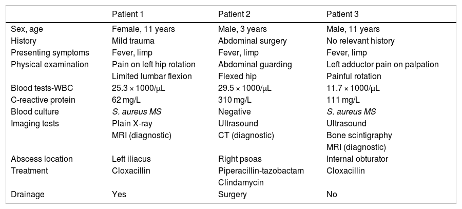
Clinical characteristics, diagnosis and treatment of presented cases.
| Patient 1 | Patient 2 | Patient 3 | |
|---|---|---|---|
| Sex, age | Female, 11 years | Male, 3 years | Male, 11 years |
| History | Mild trauma | Abdominal surgery | No relevant history |
| Presenting symptoms | Fever, limp | Fever, limp | Fever, limp |
| Physical examination | Pain on left hip rotation | Abdominal guarding | Left adductor pain on palpation |
| Limited lumbar flexion | Flexed hip | Painful rotation | |
| Blood tests-WBC | 25.3 × 1000/μL | 29.5 × 1000/μL | 11.7 × 1000/μL |
| C-reactive protein | 62 mg/L | 310 mg/L | 111 mg/L |
| Blood culture | S. aureus MS | Negative | S. aureus MS |
| Imaging tests | Plain X-ray | Ultrasound | Ultrasound |
| MRI (diagnostic) | CT (diagnostic) | Bone scintigraphy | |
| MRI (diagnostic) | |||
| Abscess location | Left iliacus | Right psoas | Internal obturator |
| Treatment | Cloxacillin | Piperacillin-tazobactam | Cloxacillin |
| Clindamycin | |||
| Drainage | Yes | Surgery | No |
CT, computed tomography; MRI, magnetic resonance imaging; MS, methicillin-sensitive; WBC, white blood cells.
Pyomyositis usually involves the muscles of the pelvis and lower extremities. Its pathophysiology is still under debate; it has been hypothesised that the association of bacteraemia and traumatic injury of the muscle may activate the inflammatory cascade that leads to abscess formation.2 In our series, patient 1 reported a history of mild trauma a few days before onset and patient 2 had undergone surgery for correction of an undescended right testicle and umbilical hernia 20 days before. Some authors describe pyomyositis as a progression through 3 stages, starting with diffuse inflammation and ending with sepsis and abscess formation. S. aureus is identified as the causative agent in up to 90% of cases, with positive blood cultures in 40%.2 Recent studies describe an increased frequency of PVL-producing S. aureus isolates in patients with pyomyositis,1 establishing an association between the detection of these strains and pyomyositis outcomes including symptom duration, complications and need of drainage or surgery.1,3 Panton–Valentine leucocidin-producing S. aureus strains can be either methicillin-sensitive (MSSA) or methicillin-resistant (MRSA),2,3 which highlights the importance of susceptibility testing after isolation in blood culture. Another pathogen that is isolated less frequently is Streptococcus pyogenes, which is associated with greater severity and even toxic shock.4 In the case series presented here, MSSA was isolated in 2 patients from culture of blood samples collected early after the initial assessment. Testing for detection of PVL was not performed.
The diagnosis of pyomyositis may be delayed due to the depth of the involved muscles and the absence of external manifestations. The physical examination and serum markers of infection offer a low specificity for differentiation of pyomyositis from septic arthritis and osteomyelitis.4 Ultrasonography is widely available and does not involve exposure to ionizing radiation, so it is useful in the initial investigation, but its diagnostic yield varies depending on who performs it and its findings may be inconclusive.5 Magnetic resonance imaging (MRI) is the gold standard of diagnosis,4 as it allows detection of pyomyositis from the early stages of disease as well as accurate assessment of the size of the abscess and adjacent structures. In our patients, an ultrasound scan and a plain radiograph were the first-line imaging tests, with normal findings in all 3 cases, and the definitive diagnosis was made by means of MRI (patients 1 and 3) and computed tomography (CT) (patient 2).
Treatment must be selected based on the stage of disease. Accurate diagnosis followed by early initiation of empirical intravenous antibiotherapy may suffice in the initial stages of pyomyositis.2,6 However, if an abscess is detected, the most recent studies recommend percutaneous drainage prior to initiation of antibiotherapy.4 In these cases, ultrasound can be very useful to assist drainage. The selected antibiotic must be effective against S. aureus, with the choice depending on severity, abscess location, epidemiology and local bacterial drug resistance patterns. If the prevalence of MRSA is low, cloxacillin or a first-generation cephalosporin may be used as first-line treatment, with no established consensus regarding the optimal duration of treatment, which usually ranges from 3 to 6 weeks.4 If there is no improvement, ultrasound- or CT-guided drainage is indicated, followed by culture of an aspirate sample, which offers a higher diagnostic yield.6 In our series, patient 1 required percutaneous drainage due to an increase in abscess size despite improvement of symptoms and laboratory markers with intravenous cloxacillin, and the culture of the drained fluid was negative. Patient 2 underwent abdominal surgery via laparotomy due to association of the abscess in the psoas with appendicitis; Streptococcus constellatus and Bacteroides fragilis were isolated in the wound drainage culture. The third patient had a favourable outcome with conservative treatment.
In conclusion, while pyomyositis is infrequent, it must be included in the differential diagnosis of fever accompanied by limping. The gold standard imaging test is MRI, and early initiation of empirical antibiotherapy can prevent complications, although drainage is required if abscesses develop or there is no improvement with antibiotherapy. Testing for detection of the PVL toxin may help predict the course of disease and the development of complications.
Please cite this article as: López Fernández L, Jiménez Escobar V, Sáenz Moreno I, Gallinas Maraña E, Cuadrado Piqueras L. Piomiositis aguda: diagnóstico y tratamiento de 3 casos en un hospital de segundo nivel. An Pediatr (Barc). 2021;95:467–468.
