
Hepatocyte nuclear factor-1 beta (HNF-1β; genetic locus 17q12) is involved in the development of several tissues in the organogenesis of the pancreas, liver and genitourinary system. Maturity-onset diabetes of the young (MODY) and renal cysts1 are the diseases in which the role of HNF-1β is well known, but its involvement in other congenital anomalies of the urinary tract is not well understood. These disorders follow an autosomal dominant pattern of inheritance. We present the case of a family affected by a variant of the HNF1B gene in which the index case was an infant that received an antenatal diagnosis of hydronephrosis manifesting with renal pelvis dilatation.
The patient was an infant aged 12 months followed up from month 1 post birth due to antenatal diagnosis of hydronephrosis with dilatation of the left renal pelvis and calyces categorised as grade 3–4 (Society for Fetal Urology classification).2 The postnatal follow-up ultrasound scan confirmed the dilatation of the renal pelvis and calices, but also detected an abnormally small left kidney (2.5 SD below mean) with 2 cortical cysts, with a normal contralateral kidney. The personal history was otherwise unremarkable. The voiding cystourethrogram was normal and the 99mTc-MAG3 diuretic renogram found normal function in both kidneys and no evidence of obstruction. The follow-up ultrasound scan at 1 year post birth evinced kidneys of normal size other than the left-sided dilatation, but with bilateral hyperechogenicity and renal cysts, while laboratory tests detected an elevated creatinine level (0.6 mg/dL) and a decreased glomerular filtration rate (GFR, Schwartz formula [2009]: 52.9 mL/min/1.73 m2). The only relevant finding of the family history was the presence of type 1 diabetes (T1D) in the father and paternal grandfather. A sister aged 3 years was healthy with normal renal sonographic features. We reviewed the medical history of the father and found that at the time of diagnosis of T1D at age 16 years, the ultrasound examination of the kidneys had found bilateral hyperechogenicity and bilateral renal cysts. Furthermore, 5 years after the diagnosis of T1D, the father had received a diagnosis of diabetic nephropathy with stage 3A chronic kidney disease (CKD) that was atypical due to the absence of albuminuria (category A1) and to adequate metabolic control (glycated haemoglobin <6.5%). Also salient was the presence of high creatinine levels from age 10 years (1.1 to 1.6 mg/dL) predating the diagnosis of DM1. The clinical picture of the grandfather was similar to that of the father.
Given the suspicion of renal cysts and diabetes syndrome in the family, we ordered testing of the HNF1B gene in the index patient, which identified a heterozygous variant (change in HNF1B, NM_000458.2: c.554 + 3_554 + 6del). Genetic testing was then performed in the father, with detection of the same mutation. At present, the patient is aged 25 months and has CKD stage G3A A1 (GFR, Schwartz formula [2009], 59 mL/min/1.73 m2 and cystatin C-Pottel equation [2017], 52 mL/min/1.73 m2) with hyposthenuria in absence of the albuminuria, metabolic acidosis, electrolyte imbalance, hypomagnesaemia, hyperuricaemia or hypertransaminasaemia characteristic of CKD. The patient is managed exclusively with dietary measures.
Changes in the HNF1B gene were responsible for the syndrome manifesting with renal cysts and diabetes, which would be categorised as an autosomal dominant tubulointerstitial kidney disease (Table 1).3 These variants typically present with renal cysts and diabetes syndrome (MODY type 5), usually with postpubertal onset. The severity of renal involvement varies widely, ranging from foetal death due to prenatal kidney failure to normal renal function in adulthood, with no phenotype-genotype correlation.1,3 They are also one of the most common causes of hyperechoic kidneys in the foetus and the most frequently identified monogenic cause of congenital anomalies of the urinary tract (prevalence of 10% to 30% depending on the case series), and may manifest with renal agenesis, polycystic kidney, unilateral or bilateral renal dysplasia, renal ectopia, vesicoureteral reflux, pyeloureteral junction stenosis or hydronephrosis,4–6 as occurred in our patient. Besides diabetes (including gestational diabetes), the most frequent extrarenal manifestations are hypomagnesemia, hypokalaemia, hyperuricemia, gout and transaminase elevation. Given that the initial presentation of a HNF1B variant may be a congenital anomaly of the kidney and urinary tract (CAKUT) and that changes in this gene are among the most frequent causes of sonographic abnormalities in the antenatal period, recommendations have been published to determine the indication of testing of the HNF1B gene in patients with CAKUT based on a series of eligibility criteria (Table 2).4,5 Awareness of this disease is important not only for the purpose of early diagnosis (to ensure adequate follow-up in these patients, improve long-term outcomes and slow down the progression of renal disease), but also to provide genetic counselling to affected families, which is particularly necessary in the case of HNF1B gene variants due to their broad phenotypic spectrum, since there may be individuals with onset of CKD in the first year of life whose parents have milder forms of disease, as was the case of the patient presented in this article.
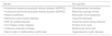
Diseases presenting with renal cysts.1
| Genetic | Non-genetic |
|---|---|
| • Autosomal recessive polycystic kidney disease (ARPKD) | Developmental anomalies |
| • Autosomal dominant polycystic kidney disease (ADPKD) | Medullary sponge kidney |
| • Nephronophthisis | Multicystic renal dysplasia |
| • Medullary cystic kidney disease | Acquired diseases |
| • DNF1β-related disease | Acquired cystic kidney disease |
| • Von Hippel-Lindau disease | Simple renal cysts |
| • Tuberous sclerosis complex | Multilocular renal cyst |
| • Renal cysts in malformation syndromes | Hypokalaemic cystic disease |
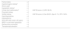
HNF1B score.5
| Family history | + 2 | |
| Hyperechogenic kidneya | + 4 | |
| Renal cystsa | + 4 | |
| Hypoplastic kidneya | + 2 | |
| Urinary tract malformationa | + 1 | HNF1B score < 8, NPV 99.4% |
| Polycystic kidney | + 2 | |
| Solitary kidney | + 1 | HNF1B score ≥ 8, Sen 98.2%, Spe 41.1%, PPV 19.8% |
| Hypomagnesemia | + 2 | |
| Hypokalaemia | + 1 | |
| Gout with early onset (<30 years) | + 2 | |
| MODY or pancreas hypoplasia | + 4 | |
| Genitourinary anomalies | + 4 | |
| Transaminase elevation | + 2 |
MODY, maturity-onset diabetes of the young; NPV, negative predictive value; PPV, positive predictive value; Sen, sensitivity; Spe, specificity.
Please cite this article as: Alarcón-Alacio MT, Penela-Vélez de Guevara MT, Ballesteros-García MM, Rivero-Martín MJ. Ectasia piélica antenatal: signo guía para el diagnóstico familiar de una enfermedad genética. An Pediatr (Barc). 2021;95:204–206.
