


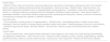

Cholestasis is indicative of hepatobiliary dysfunction and is always pathological. Early detection helps improve the prognosis of some of the underlying diseases that cause it. The most common liver disease that causes cholestasis in the first months of life is biliary atresia, followed by monogenic diseases. The objective of this document is to provide consensus-based recommendations for the adequate management of cholestasis based on the review of the current evidence. A working group was created for the purpose, with participation of members of the Spanish Society of Pediatric Gastroenterology, Hepatology and Nutrition, the Spanish Association of Primary Care Pediatrics and the Spanish Society of Primary Care Pediatrics. The group established 26 recommendations to guide management in everyday clinical practice in both primary care and hospital settings.
La colestasis indica disfunción hepatobiliar y es siempre patológica. Su detección precoz contribuye a mejorar el pronóstico de algunas enfermedades subyacentes que la provocan. La hepatopatía más común que causa colestasis en los primeros meses de vida es la atresia de vías biliares, seguida por enfermedades genéticas monogénicas. El objetivo de este documento es establecer un consenso para un adecuado manejo de la colestasis mediante revisión de la evidencia disponible. Para ello, se constituyó un grupo de trabajo con participación de miembros de la Sociedad de Gastroenterología, Hepatología y Nutrición Pediátrica, la Asociación Española de Pediatría de Atención Primaria y la Sociedad Española de Pediatría de Atención Primaria. Se establecieron 26 recomendaciones con el objetivo de que sirvan de utilidad en la práctica clínica habitual, tanto en atención primaria como hospitalaria.

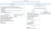
Cholestasis is indicative of hepatobiliary dysfunction and, in the pediatric population, is uncommon but potentially serious. Cholestasis affects approximately one in 2500 newborn infants, in whom the most frequent cause is biliary atresia.1 Early diagnosis is crucial for its correct management and to improve patient outcomes. The aim of this document is to establish guidelines for the appropriate diagnosis and management of cholestasis, which were developed by consensus by primary care pediatricians and specialists in pediatric gastroenterology, hepatology and nutrition (Fig. 1).
Methods
The first step was the formation of a working group composed of seven members representing the Sociedad de Gastroenterología, Hepatología y Nutrición Pediátrica (Society of Gastroenterology, Hepatology, and Pediatric Nutrition), the Asociación Española de Pediatría de Atención Primaria (Spanish Association of Primary Care Pediatrics), and the Sociedad Española de Pediatría Extrahospitalaria y Atención Primaria (Spanish Society of Outpatient Pediatrics and Primary Care). The group formulated twelve clinical questions regarding the detection, focused diagnostic evaluation and management (nutritional, pharmacological, and surgical) of cholestasis.
The group conducted a literature search in PubMed for “pediatric cholestatic liver disease” restricted to sources published between January 2015 and December 2023 and to ages 0–18 years and for “genetic liver disease” and “histology liver disease” with no age or time restrictions. We used the shared Zotero 5.0 reference management tool. The search yielded 446 articles published in English or Spanish for which we consulted the full text. Other articles considered pertinent by reviewers for answering the formulated questions were added to the search results. Of the total articles considered, 90 were considered relevant for the development of the guideline and selected because they were suitable for answering the clinical questions that the group had formulated for the consensus process. The excluded articles either did not address the pediatric population, covered topics unrelated to cholestasis or had methodological limitations.
Development of the documentEach clinical question was answered by one member of the group (as appropriate based on the expertise of each member) based on the available evidence. The group did not carry out a systematic review or meta-analysis of the evidence. All members of the group reviewed all the answers a total of four times (using Google Drive), and an online meeting was held after each of these review rounds (using Microsoft Teams). All the recommendations in this guideline were established by consensus and approved by the members of the group (informal consensus process). An online vote (Google Forms) was held to rate each recommendation on a six-point Likert scale (1: strongly disagree; 2: disagree; 3: somewhat disagree; 4: somewhat agree; 5: agree; 6: strongly agree). Recommendations were approved once consensus was reached by more than 80% of participants (with ratings of 5–6). The full consensus document is available for consultation at the webpages of each of the participating scientific societies.
Cholestasis at the biochemical levelCholestasis is defined as reduced bile formation or flow that may manifest with jaundice (yellowish discoloration of skin, sclera and other tissues resulting from the accumulation of excess bilirubin), partial (hypocholia) or total (acholia) discoloration of feces, choluria (dark-colored urine due to the presence of bilirubin) and pruritus.1–3
At the biochemical level, it is defined as elevated direct/conjugated bilirubin levels (serum bilirubin > 1 mg/dL or direct fraction > 20% of total bilirubin), usually accompanied by elevation of serum bile acids. Other markers of cholestasis are elevation of the enzymes gamma-glutamyl transferase (GGT) and alkaline phosphatase (AP) and of substances usually excreted in bile, such as cholesterol.1–3
Reference values for GGT vary with age, and its secretion can be induced by alcohol and drugs, which complicates its interpretation.4 Normal ranges in the healthy pediatric population have not been clearly established.4–8 Adeli et al. proposed the following upper limits of normal stratified by age in the healthy population: 219 U/L in neonates between days 0–15 post birth; 127 U/L in infants aged 15 days to 1 year; 20 U/L from 1 to 18 years.5
Elevation of AP can be difficult to interpret in children, as it is frequently elevated on account of increases in bone isoenzyme levels.9 Consequently, GGT is a more specific marker of cholestasis than PA in the pediatric population.4,9
Recommendations- 1
We recommend the use of the following upper limit of normal values for GGT, stratified by age: 219 U/L between days 0–15 post birth; 127 U/L in infants aged 15 days to 1 year; 20 U/L from 1 to 18 years.
- 2
We suggest interpreting AP values in the context of all other markers of liver injury.
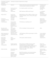
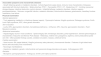
The etiology of cholestasis varies with age. Newborn infants have immature livers predisposing to the development of cholestasis. In this age group, cholestasis is usually transient and resolves along with the underlying condition that caused it (prematurity, severe hypoxia, heart disease, sepsis, surgery, intestinal failure or prolonged parenteral nutrition). In the absence of predisposing factors, cholestasis may result from multiple intrahepatic and extrahepatic causes (Table 1).1,2,10,11 The most frequent cause is biliary atresia (BA), responsible for 25% to 40% of cases, followed by genetic/metabolic disorders (eg, progressive familial intrahepatic cholestasis [PFIC], alpha-1 antitrypsin deficiency).10,12,13
Causes of cholestasis in neonates and infants and associated genes.
| Disease | Gene | Disease | Gene | Disease | Gene |
|---|---|---|---|---|---|
| Extrahepatic causes | Tight junction defects | Lysosomal storage disease | |||
| Biliary atresia Choledochal cyst Cholelithiasis, choledocholithiasis Inspissated bile/mucous plug Congenital perforation of the common bile duct | – – – – | TJP2 deficiency (PFIC4) | TJP2 | Niemann-Pick type C | NPC1; NPC2 |
| Intrahepatic causes | USP53 deficiency (PFIC7) | USP53 | Niemann-Pick type A and B | SMPD1 | |
| Infections | NISCH syndrome | CLDN1 | Lysosomal acid lipase deficiency (Wolman disease) | LIPA | |
| Congenital (TORCH) or postnatal: CMV, herpes virus 1-2-6, toxoplasmosis, rubella, parvovirus B19, enterovirus (coxsackie, echovirus), adenovirus, syphilis, HIV, Listeria monocytogenes, SARS-CoV-2, congenital TB | – | Bile acid synthesis and conjugation disorders | Gaucher disease | GBAPSAP (encodes its activator protein, saposin C) | |
| Toxic and secondary cholestasis | 3-β-HSD-oxidoreductase deficiency, CBAS1 | HSD3B7 | Mitochondrial disorders | ||
| Parenteral nutrition associated cholestasis (PNALD); drugs (ceftriaxone, erythromycin, rifampicin, furosemide); intestinal obstruction; cardiovascular disorders; neoplastic disorders; perinatal asphyxia Liver immaturity (preterm birth) | – | D4-3-oxosteroid 5 β-reductase deficiency, CBAS2 | AKR1D1 | Mitochondrial DNA depletion syndrome | POLG, DGUOK, MPV17 |
| Cholestasis with involvement of other organs | Oxisterol-7α-hydroxylase deficiency | CYP7B1 | SUCLG1, C10ORF2, elongation factor G1,TRMU related and BCS1L deficiency | SUCLG1, C10ORF2, EGF1, TRMU, BCS1L | |
| α-1-antitrypsin deficiency | SERPINA1 | 2-methylacil-CoA racemase deficiency, CBAS4 | AMACR | Mitochondrial Fatty Acid Oxidation defects | |
| Cystic fibrosis | CFTR | BAAT deficiency | BAAT | LCHAD/MTP deficiency | HADHA, HADHB |
| Defects of the biliary canalicular transport (bile acids or phospholipids) | BACL deficiency | SLC27A5 | Peroxisomal disorders | ||
| PFIC1; FIC1 deficiency | ATP8B1 | Cerebrotendinous xanthomatosis | CYP27A1 | Zellweger spectrum disorders | PEX genes |
| PFIC2; BSEP deficiency | ABCB11 | Biliary development defects | Aminoacidopathies | ||
| PFIC2; MDR3 deficiency | ABCB4 | Alagille syndrome | JAG1, NOTCH 2 | Primary bile acid malabsorption | SLC51B |
| PFIC5; FXR deficiency | NR1H4 | Neonatal sclerosing cholangitis | DCDC2 | Biliary, renal, neurologic, and skeletal syndrome.; OMIM 619534 | TTC26 |
| PFIC6 | MYO5B | Neonatal ichthyosis-sclerosing cholangitis | CLDN1 | Carbohydrate metabolism defects | |
| Larsen syndrome | LRS | Arthrogryposis renal dysfunction cholestasis syndrome (ARC) | VPS33B (VIPAR), VIPAS39 | Classic galactosemia | GALT |
| PFIC8; KIF12 deficiency | KIF12 | Caroli disease | PKHD1 | Glycogen storage disease type IV | GBE1 |
| Ciliopathies (biliary duct, PFIC9) | Several genes (ZFYVE19) | Congenital disorders of glycosylation | Different genes | ||
| Hematologic and immune-mediated disorders | Inborn error of polyols and pentose metabolism | Syndromic cholestasis | |||
| Hemophagocytic lymphohistiocytosis | Different genes, such as PRF1, UNC13D, STX11, STXBP2, RAB 27, XL | Down and Edwards syndrome | Trisomy 21 and 18 | ||
| Neonatal hemochromatosis (GALD and non-GALD) | Non-GALD Eg: DGUOK, SRD5B1, BCS1L | Transaldolase deficiency | TALDO111 | Kabuki syndrome | KMT2D, KDM6A, MLL2 |
| Congenital lupus | – | Cellular trafficking abnormalities | Noonan syndrome | PTPN11, SOS1, RAF1 and KRAS | |
| Posthemolytic cholestasis | – | NBAS deficiency | NBAS | Aagenaes syndrome | LSC1, CCBE1 |
| Cholesterol metabolism disorders | CALFAN syndrome (cholestasis, acute liver failure, and neurodegeneration) | SCYL1 | Endocrine disorders | ||
| Smith-Lemli-Opitz syndrome | DHCR7 | Other | Thyroid disorder | Different genes in CHT; eg, FOXE1, NKX2-1/5, PAX8, SLC26A4, TSHR | |
| Mevalonic aciduria | MVK | SLC51A deficiency | SLC51A | [1,0]Panhypopituitarism | Different genes in genetic forms |
| Urea cycle defects | Primary bile acid malabsorption | SLC51B | Eg: HESX1, PROP1, POUF1, LHX3, LHX4, GLI2, SOX3 | ||
| Urea cycle defects | OTC, ASS, ASL; ARG | Biliary, renal, neurologic, and skeletal syndrome, OMIM 619534 | TTC26 | [1,0]Adrenal insufficiency | Monogenic, eg: POR, MC2R, MRAP, StAR, AYP11A1, NNT, TRXR2 |
| Citrin deficiency (NICCD) | SLC25A13 | MEDNIK syndrome | AP1S1 | Syndromic, eg CDKN1C, MCM4, SAMD9, SGPL1 | |
Abbreviations: CBAS, congenital bile acid synthesis disorder; CMV, cytomegalovirus; GALD, gestational alloimmune liver disease; HIV, human immunodeficiency virus; MEDNIK, mental disability, enteropathy, deafness, neuropathy, ichthyosis, and keratoderma; PFIC, progressive familial intrahepatic cholestasis; TB, tuberculosis; TORCH, toxoplasmosis, other, rubella, cytomegalovirus and herpes simplex.
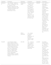
The causes of cholestasis in older children (Table 2) can be classified in two groups according to the presence or absence of transaminitis.2,14
Causes of cholestasis in children.
| Extrahepatic cholestasis (anatomical biliary obstruction) | Intrahepatic cholestasis (hepatocellular damage) | ||
|---|---|---|---|
| Cholelithiasis | Infections | Defects of the biliary canalicular transport (bile acids or phospholipids) and tight junction defects | Cholestasis with involvement of other organs |
| Calculous or acalculous cholecystitis | Acute and chronic viral hepatitis, sepsis | FIC1 deficiency: BRIC1 and ICP type 1 | Cystic fibrosis (CFTR3) |
| Choledochal cyst | Autoimmune | BSEP deficiency: BRIC2 and ICP type 2 | α-1-antitrypsin deficiency (SERPINA 1) |
| Biliary stricture | Autoimmune hepatitis with or without sclerosing cholangitis | MDR3 deficiency: PFIC 3, BRIC3, ICP type 3, drug-induced cholestasis, LPAC, cirrhosis (with copper overload) | Hematological and immune-mediated |
| Primary and secondary sclerosing cholangitis | Toxic and secondary | TJP2 deficiency: ICP | Graft-vs-host disease (bone marrow transplant) |
| Portal biliopathy | Toxins (natural remedies) and pharmaceuticals (paracetamol, isoniazid, valproic acid) | FXR deficiency: cholelithiasis, ICP | Hemophagocytic syndrome |
| Bile duct cancer (rhabdomyosarcoma, cholangiocarcinoma) | Liver disease secondary to intestinal failure and prolonged parenteral nutrition | PFIC 6: cholestasis in first two years of life | Other |
| Biliary obstruction by a tumor (lymphoma), tuberculosis | Heart failure and reduced blood flow | USP53 deficiency: BRIC-like phenotype in children and adults | Tumor infiltration: primary hepatic lymphoma or metastases |
| IgG4-related autoimmune pancreatitis associated with sclerosing cholangitis | Biliary development defects | Other genetic-metabolic disorders | Hypothyroidism |
| AIDS cholangiopathy | Alagille syndrome (JAG1, NOTCH 2) | Wilson disease (ATP7B), tyrosinemia (FAH), hereditary fructose intolerance (ALDOB), mitochondrial disorders (POLG, DGUOK, MPV17, SUCLG1, C10ORF2, EGF1, TRMU, BCS1L), Niemann-Pick Type C (NPC1, NPC2) | Normal liver function (isolated conjugated hyperbilirubinemia) |
| Biliary ascariasis | Nonsyndromic bile duct paucity | Vascular anomalies | Dubin-Johnson syndrome (ABCC2 gene) |
| Autosomal recessive polycystic kidney disease (PKHD1, DZIP1L genes) | Budd-Chiari syndrome, veno-occlusive disease (history of chemotherapy), portal biliopathy, hemangiomas | Rotor syndrome (SLC01B1, SLC01B3 genes) | |
Abbreviations: BRIC, benign recurrent intrahepatic cholestasis; BSEP: bile salt export pump; FXR: farnesoid X receptor; ICP, intrahepatic cholestasis of pregnancy; LPAC, low phospholipid-associated cholelithiasis syndrome; MDR3: multidrug resistance protein 3; TJP2: tight junction protein 2; USP53, ubiquitin-specific peptidase 53.
(Expansions provided for proteins, but not for genes).
- 1
We recommend an etiological assessment based on the age and circumstances of the patient, including intrahepatic and extrahepatic causes in the differential diagnosis.
The most frequent causes of non-cholestatic jaundice in infants are physiologic jaundice and a prolonged form in breasted infants known as breast milk jaundice.
In contrast, cholestatic jaundice is always pathological, and its etiology needs to be investigated. Early diagnosis of causes that can be treated is essential, for instance, BA, in which early surgical intervention (first 6 weeks) have a positive impact on patient outcomes.15
The role of the primary care pediatrician is crucial in the early detection of cholestatic jaundice. Therefore, cholestasis should be ruled out in all newborns with prolonged jaundice (beyond 15 days post birth). If the infant is breastfed and there are no red flags (weight faltering, acholia, choluria, visceromegaly and/or abdominal distension), international guidelines recommend performance of blood tests (total and direct bilirubin) at 3 weeks of age if jaundice persists.1
Recommendations- 1
Rule out cholestasis by measuring total and conjugated bilirubin levels in any infant with jaundice persisting beyond 15 days post birth in formula-fed infants or 3 weeks post birth in breastfed infants without warning signs.
- 2
In the presence of warning signs, we recommend performance of blood tests on an urgent basis, regardless of the age of the infant.
- 3
We do not recommend the use of transcutaneous jaundice meters for screening of cholestasis in infants.
There are three key elements in the evaluation of a patient with suspected cholestasis: a detailed history-taking, a thorough physical examination and liver function tests.1,16 In the history-taking, emphasis should be placed on the prenatal, perinatal and postnatal history of the patient and the family history (Table 3).16 The most characteristic signs and symptoms of cholestasis are jaundice, acholia or hypocholia, choluria and pruritus, although not all of them are present in every case (Table 4).1 Hypocholia may be underestimated,1,17 so the use of stool color charts1 or mobile applications (eg, PopòApp) is recommended for its assessment.
Aspects that need to be included in the history-taking of a patient with cholestasis.
| Family history |
| - Obstetric history: history and outcomes of previous pregnancies, spontaneous miscarriage or siblings who died in the neonatal period, exposure to substances/pharmaceuticals during gestation, maternal serology - Prenatal history: maternal pruritus, liver dysfunction, gestational cholestasis, fever or lymphadenopathy during pregnancy - Cholestasis in other family members (cystic fibrosis, α-1-antitrypsin deficiency, progressive familial intrahepatic cholestasis, Alagille syndrome) - Other relevant hepatic or extrahepatic diseases in other family members (especially those manifesting with hemolysis or cardiac or vascular involvement) - Consanguinity (increased risk of genetic or metabolic disorders) |
| Neonatal history |
| - Use of medication (including vitamin K supplementation) - APGAR scores - Infant feeding/nutrition in relation to the onset of cholestasis. Particular interest in knowing whether parenteral nutrition was needed - Delayed passage of meconium (cystic fibrosis) - Gestational age and birth weight - Neonatal infection - Heel prick test - Need for surgery (necrotizing enterocolitis, intestinal atresia) |
| Current history |
| - Timing of jaundice onset - Stool pigment (direct observation of stool pigment or use of stool color charts strongly recommended) and appearance (steatorrhea) - Color and odor of urine - Presence of other symptoms: pruritus, vomiting, weight faltering, irritability, psychomotor delay, asthenia, anorexia, abdominal pain, hearing and/or vision impairment - Presence of other known diseases |
Associated signs and symptoms to assess for in patients with cholestasis.
| Associated symptoms and characteristic diseases |
| - Growth faltering: genetic or metabolic disorders - Acholic/hypocholic (pale) stools, choluria: more characteristic of diseases associated with bile duct obstruction - Watery diarrhea: PFIC1 - Pancreatitis: PFIC1, CF - Steatorrhea: CF - Vomiting: lysosomal storage disease, intestinal obstruction, pyloric stenosis - Irritability/lethargy: metabolic disorders, infection (sepsis), panhypopituitarism - Seizures: congenital infection, metabolic disorders, mitochondrial diseases, galactosemia, hereditary fructose intolerance, liver failure |
| Physical examination |
| General appearance |
| - Toxic-appearing: metabolic or infectious disease (sepsis) - Dysmorphic features: Alagille syndrome, Zellweger syndrome, Smith-Lemli-Opitz syndrome, ARC, other genetic disorders |
| Skin evaluation |
| - Laxity: ARC, glycosylation disorders, transaldolase deficiency - Ichthyosis: ARC, Gaucher, glycosylation disorders - Rash: Mevalonic aciduria |
| Abdominal examination |
| - Hepatomegaly: found in most conditions - Splenomegaly: BA, hematologic disorders, portal hypertension. Severe splenomegaly is characteristic of Gaucher and Nieman Pick disease - Heterotaxy, midline liver, polysplenia, asplenia, and/or preduodenal portal vein: BA - Other possible findings: ascites, collateral circulation, masses, umbilical hernia |
| Cardiac examination |
| - Heart murmur: BA (septal defects), Alagille (pulmonary artery stenosis), chromosomal disorder - Features of right-sided heart failure (with secondary liver involvement) |
| - Neurologic manifestations |
| - Hypotonia: metabolic, genetic, mitochondrial and lysosomal storage disorders and sepsis - Arthrogryposis: ARC |
| Genitalia |
| - Micropenis: panhypopituitarism - Ambiguous: Smith-Lemli-Opitz syndrome |
Abbreviations: ARC, arthrogryposis-renal dysfunction-cholestasis syndrome; BA, biliary atresia; CF, cystic fibrosis; PFIC, progressive familial intrahepatic cholestasis.
The physical examination should be thorough and include a body systems review,1,2 assessing for the presence of hepatomegaly, splenomegaly, other signs of portal hypertension and faltering growth, among others (Table 4).14
As regards laboratory tests, the initial diagnostic evaluation should include total and conjugated bilirubin measurement and a full liver function panel. Unless physiologic or breast milk jaundice are suspected, it is also indicated to order first-tier tests for etiological diagnosis in the initial workup, as cholestasis requires an expedited focused investigation.
Recommendations- 1
The following are recommended for the initial evaluation of a patient with suspected cholestasis: a detailed history-taking, a thorough physical examination with body system review and laboratory tests for biochemical markers of cholestasis.
- 2
In infants with suspected cholestasis, the use of stool color charts is recommended to assist providers in the assessment of acholia/hypocholia.
- 3
We propose the inclusion of a stool color chart in the child health booklet issued by the public health system to allow identification of hypocholia/acholia by caregivers.
The differential diagnosis of the various conditions that cause cholestasis can be complex. In infants with confirmed cholestasis, early diagnosis of diseases for which specific treatments are available is a priority. The evaluation should be carried out in stages based on the suspected diagnosis to determine the etiology while assessing the severity of liver disease (Fig. 1). The causes of cholestasis in older children differ significantly from those in infants, so the diagnostic evaluation will be different in these age groups (Fig. 1).
All patients with cholestasis should undergo an initial diagnostic evaluation including a full liver function panel (alanine aminotransferase, aspartate aminotransferase, GGT, AP, total/conjugated bilirubin, glucose, albumin, coagulation). In infants, it is also important to review the newborn screening results.1
Imaging tests are particularly important to rule out anatomical causes of cholestasis.3Abdominal sonography (following a fast of at least 4 h) is a simple, noninvasive technique that is useful as a first step to rule out obstructive causes, lesions of the biliary tree or choledochal cysts. Several sonographic findings are suggestive of biliary atresia, although none allows definitive diagnosis.18Intraoperative cholangiography is considered the gold standard for diagnosis of biliary atresia, so it should not be delayed if the condition is strongly suspected.3
Recommendations- 1
When cholestasis is suspected, we recommend a full liver function panel and imaging by means of sonography, with subsequent tests selected based on patient age and the suspected diagnosis (tests for infection, metabolic study, imaging, genetic testing, liver biopsy).
- 2
In the case of suspected biliary atresia, we recommend an evaluation based primarily on abdominal sonography followed by intraoperative cholangiography. The latter should not be delayed while awaiting the results of other diagnostic tests.
Early detection and etiological diagnosis of cholestasis are of utmost importance, so, once cholestasis has been confirmed, we recommend timely referral for assessment by a pediatric gastroenterologist/hepatologist.1
Under certain circumstances (cholestasis in an infant, clinical or laboratory evidence of associated liver failure, hemodynamic and/or respiratory instability, or suspected neoplastic disease), the patient should be admitted to a referral hospital for immediate evaluation.19
Recommendations- 1
Hospital admission for urgent evaluation is recommended in any of the following circumstances: cholestasis in an infant, findings suggestive of liver failure, hemodynamic and/or respiratory instability, or suspected neoplastic disease.
- 2
In the case of cholestasis in a child (no longer an infant), we recommend urgent referral for assessment by a pediatric gastroenterologist/hepatologist.
It is estimated that between 25% and 50% of cases of cholestasis are due to identifiable genetic changes. These involve a broad range of genes that have a direct or indirect effect on bile synthesis, transport and flow. Once surgical (AVB, etc.), infectious and secondary causes have been ruled out, cholestasis in children is most likely due to a monogenic liver disease (Tables 1 and 2).10 The geneticist plays a key role in determining the most suitable genetic test for each case.
Recommendations- 1
After ruling out anatomical, viral and metabolic causes, we recommend genetic testing for monogenic causes of cholestasis.
- 2
We recommend performing genetic testing after agreeing with the geneticist on the best option for the patient (next-generation sequencing, clinical exome, whole exome, etc).
Advances in noninvasive diagnostic methods (blood chemistry tests, imaging techniques and genetic analysis) have changed the diagnostic use of liver biopsy to a secondary level.20,21 A liver biopsy should be performed in cases in which the findings of previous tests have not allowed identification of specific etiology and cases in which the degree of hepatic involvement needs to be determined more precisely.1,10Table 5 presents the possible biopsy findings in different diseases.
Liver biopsy: histological findings in the main causes of cholestasis.
| Disease | Findings |
|---|---|
| Extrahepatic biliary atresia | Bile duct proliferation, bile plugs, portal stromal edema |
| Alagille syndrome | Intrahepatic bile duct paucity (this may not be evident in young infants) |
| Progressive familial intrahepatic cholestasis | |
| Type 1 (FIC 1 deficiency) | Ductopenia |
| Type 2 (BSEP deficiency) | Giant cell transformation |
| Type 3 (MDR3 deficiency) | Ductal proliferation with fibrosis |
| Inborn error of bile acid synthesis | Nonspecific: inflammation without bile duct proliferation, giant cell transformation |
| α-1-antitrypsin deficiency | Eosinophilic globules, PAS-positive and diastase-resistant hepatocyte inclusions |
| Congenital panhypopituitarism | Giant cell hepatitis with bile duct hypoplasia |
| Autoimmune hepatitis | Interface hepatitis, lobular hepatitis or bridging necrosis. |
| Wilson disease | Steatosis, inflammation, fibrosis and cirrhosis. Liver copper concentration (dry weight) > 250 µg/g |
| Drug-induced liver injury | Cholestasis, hepatitis, fibrosis and inflammation; plasma cell infiltration in some cases |
Abbreviations: BSEP, bile salt export pump; MDR3, multidrug resistance-associated protein 3; PAS, Periodic Acid-Schiff stain.
- 1
Performance of a liver biopsy is recommended when it can provide additional diagnostic and/or prognostic information about the cholestatic disease.
- 2
The decision to perform a liver biopsy should not delay surgical exploration of the bile duct if indicated.
Patients with cholestasis often have associated symptoms and complications secondary to malnutrition, including growth faltering, fat-soluble vitamin deficiency and metabolic bone disease, among others. Malnutrition is common and has a multifactorial etiology in these patients,22 who also exhibit a 30% increase in the basal metabolic rate.11,23 In the presence of portal hypertension, the congestion of the intestinal mucosa causes malabsorption.22
On the other hand, the decreased concentration of bile salts in the bowel results in fat malabsorption.11,13 This can lead to fat-soluble vitamin deficiency, which carry a risk of coagulopathy, rickets and defective neurological, immunological and visual functions.24
When assessing nutritional status in these patients, it must be taken into account that body weight and associated measures may lead to underdiagnosis of acute malnutrition, as they are affected by the presence of ascites, edema or visceromegaly.25,26 The brachial or mid-upper arm circumference is a possible alternative for assessing short-term nutritional status and has been validated as a marker of malnutrition in patients with chronic cholestasis.26
In addition to the customary laboratory tests for assessment of nutritional status, fat-soluble vitamin levels should be monitored in these patients (Table 6).11,24,27
Main available multivitamin supplements and their composition.
| Preparation | Composition | Preparation | Composition | Preparation | Composition |
|---|---|---|---|---|---|
| Hidropolivit ® | 1 mL = 28 drops: - Vitamin A: 1500 IU - Cholecalciferol: 600 IU - Vitamin E 10 mg - Riboflavin (Vitamin B2) 2 mg - Pyridoxine (Vitamin B6) 1.6 mg - Ascorbic acid (Vitamin C) 50 mg - Biotin 0.125 mg - Nicotinamide 12.5 mg | Hidropolivital baby® | In 30 drops (1 mL): - Vitamin A: 400 µg - Thiamine (Vitamin B1): 0.6 mg - Riboflavin (Vitamin B2): 0.9 mg - Pantothenic acid (Vitamin B5): 6 mg - Vitamin B6: 0.6 mg - Vitamin B12: 0.7 µg - Vitamin C: 12 mg - Vitamin D: 10 µg (equivalent to 400 IU) - Zinc: 4 mg | FIADEK® | 1 mL: - Vitamin A (β-carotene 100%) 2500 IU - Vitamin D3 (cholecalciferol) 1000 IU - Vitamin E (D-alpha-tocopherol) 50 IU (33.33 mg) - Vitamin K1 (phytomenadione) 200 mg - Vitamin C (ascorbic acid) 40 mg - Vitamin B1 (thiamine) 0.5 mg - Vitamin B2 (riboflavin) 0.5 mg - Vitamin B3 (niacin) 6 mg - Vitamin B6 (pyridoxine) 0.6 mg - Vitamin B8 (biotin) 15 µg - Vitamin B5 (pantothenic acid) 3 mg - Zinc 0.39 mg - Selenium 4.4 mg |
| DEKAs Essential® | 1 mL: - Vitamin D3: 2000 IU - Vitamin E (tocofersolan): 75 IU (50 mg) - Vitamin K: 2 mg - Vitamin A: 2500 IU | ||||
| Protovit® | 1 mL (24 drops) contains: - Retinol (Vitamin A) 3000 IU - Thiamine (Vitamin B1) 2.0 mg - Riboflavin (Vitamin B2) 1.5 mg - Nicotinamide (Vitamin PP) 15.0 mg - Pyridoxine (Vitamin B6) 2.0 mg - Dexpanthenol 10.0 mg - Propylene glycol 220.0 mg - Ascorbic acid (Vitamin C) 80.0 mg - Biotin 0.2 mg - Ergocalciferol (Vitamin D) 900 IU - DL-alpha-tocopheryl acetate (Vitamin E) 15.0 mg | DHA vit® | 1 mL: - Vitamin E: 5 mg - Vitamin A: 175 µg (583 IU) - Vitamin D3 10 µg (400 IU) - DHA (docosahexaenoic acid) 40 mg | Lunafen® | One softgel: - Vitamin A: 2664 IU - Vitamin D: 10 µg - Vitamin E: 6.7 mg - Ascorbic acid: 70 mg - Thiamine (Vitamin B1): 2.46 mg - Riboflavin (Vitamin B2): 3.4 mg - Nicotinamide (Vitamin B3): 17 mg - Pyridoxine (Vitamin B6): 3.29 mg - Cyanocobalamin (Vitamin B12): 2.2 µg - Folic acid: 0.6 mg - Iron: 30 mg - Zinc: 15 mg - Calcium: 125 mg |
Increasing energy intake is recommended (delivering about 130% of the recommended calorie intake for the patient’s ideal weight),23,25 and modifying fat intake to increasing the proportion of medium chain triglycerides (MCTs), which are absorbed by passive diffusion. Restriction of protein intake is not recommended.22 Maintaining breastfeeding is also recommended, with the possibility of breastmilk fortification and MCT supplementation.20 If the infant requires supplemental formula, a formula containing larger amounts of MCTs should be chosen, with frequent use of formulas with partially or fully hydrolyzed protein.28 Patients with cholestasis require supplementation with fat-soluble vitamins, and the need of calcium supplementation should be considered given the risk of decreased bone mineral density.25,29
Recommendations- 1
As part of the nutritional support strategy in patients with cholestasis, we recommend:
- a
Adjusting energy intake to 130% of the recommended allowance based on the ideal weight.
- b
Base fat intake on medium chain triglycerides.
- c
Do not restrict protein intake, unless specifically indicated.
- d
Monitor fat-soluble vitamin levels and supplement as needed.
- a
Pruritus is one of the most common symptoms in patients with cholestasis. It can be severe enough to be disabling. Its pathogenesis is complex, multifactorial and not well understood. It is caused by the accumulation of pruritogenic substances (bile acids, lysophosphatidic acid, etc), whose circulating levels do not necessarily correlate to the intensity of the pruritus.30 An objective measurement tool is necessary to optimize the management of pruritus. Different scales are available for assessing pruritus, such as the Itch-Ro scale and the PRUCISION scale, that have been validated in the pediatric population.
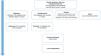
The pharmacological treatment of pruritus is symptomatic and acts through the promotion of choleresis and the modification of bile acid composition. A combination of drugs is often required to target several of the pathways involved in pruritus.31,32Fig. 2 proposes a possible regimen with the main drugs used for treatment of cholestatic pruritus.

When medical treatment fails, invasive treatment is an option in select cases, for instance, biliary diversion, which aims to reduce ileal resorption of bile acids through the surgical interruption of the enterohepatic circulation.33 Another possible treatment is albumin dialysis via the molecular adsorbent recirculating system (MARS), which allows elimination of pruritogenic substances through their binding albumin.34 Intractable pruritus can seriously impair quality of life and is, therefore, an indication for liver transplantation.
Recommendations- 1
We recommend using an pruritus measurement scale to monitor the response to treatment.
- 2
We recommend inclusion of general skin care measures and treatment with ursodeoxycholic acid in the initial management of pruritus.
- 3
If the pruritus is not controlled with these initial measures, we recommend the use of conventional drugs and/or ileal bile acid transporter (IBAT) inhibitors as indicated (see the following section).
- 4
If pharmacological treatment fails, we propose considering invasive interventions or liver transplantation.
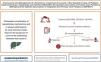
Ileal bile acid transporters (IBATs) are proteins located in the luminal surface of terminal ileal enterocytes. Their function is to mediate the resorption of bile acids, completing the enterohepatic circulation cycle. Ileal bile acid transporter inhibitors block this process and promote the fecal excretion of bile acids.35 Two molecules have been developed (odevixibat35 and maralixibat36) that are currently approved for treatment of progressive familial intrahepatic cholestasis (PFIC) and Alagille syndrome37 in children, so far limited to hospital use.
Recommendations- 1
Consider the use of IBAT inhibitors in the following scenarios:
- a
Odevixibat is approved for treatment of patients with PFIC from age 6 months to reduce the serum concentration of biliary acids and the associated pruritus.
- b
Maralixibat is approved for treatment of cholestatic pruritus in patients with Alagille syndrome from age 2 months.
- a
- 1
Flexible timing of transition based on the clinical stability, readiness and psychosocial support of the patient.
- 2
Implementation of care transition protocols involving both the pediatric and adult care teams, adapted to the particular characteristics of the center.
- 3
Assessment of mental health and social work needs of the patient.
In the pediatric age group, cholestasis is an uncommon but potentially serious condition. This consensus document provides guidance for a structured approach to management, including the early detection of signs and symptoms, the differential diagnosis and adequate treatment. Particular emphasis is placed on the early detection of treatable causes, such as biliary atresia, given the potential impact of on patient outcomes. Multidisciplinary collaboration and the use of specific diagnostic tools (e.g., genetic tests) are essential to improve the management of these patients.
FundingThis research project did not receive specific financial support from funding agencies in the public, private or not-for-profit sectors.
Maria Mercadal-Hally has occasionally collaborated in courses sponsored by IPSEN, has served as a consultant in an advisory board for IPSEN, and has received financial support to attend congresses from IPSEN and Mirum.
Inés Loverdos has occasionally collaborated in courses sponsored by IPSEN and Mirum and has served as a consultant in an advisory board for Mirum.
Ana Moreno has served as a consultant in an advisory board for Albireo.
The remaining authors declare no conflict of interest.
We thank the scientific societies that have provided support for this project (Sociedad Española de Gastroenterología, Hepatología y Nutrición Pediátrica, la Asociación Española de Pediatría de Atención Primaria and Sociedad Española de Pediatría de Atención Primaria) as well as Dr Monica Rodríguez Salas for her contribution to the original concept.
