


Hirschsprung-associated enterocolitis is a significant cause of morbidity and mortality in infants with Hirschsprung's disease. The fact that the symptoms are so variable and unspecific leads to a slow or incorrect diagnosis. The purpose of this study is to identify clinical factors associated with the diagnosis, as well as to evaluate the subsequent management of children with suspected Hirschsprung-associated enterocolitis in a paediatric emergency department.
Material and methodsA retrospective descriptive study was conducted on patients with Hirschsprung's disease who were seen in a paediatric emergency department between April 2011 and November 2015 due to clinical symptoms compatible with enterocolitis. An analytical multivariate analysis was also performed on the epidemiological and clinical variables associated to enterocolitis.
ResultsA total of 75 consultation episodes in the Paediatric Emergency Department were studied, of which 52% (39) were finally diagnosed as enterocolitis. Overall, diarrhoea was the most frequent reason for consultation (74.7%). Lethargy, abdominal distension, and pathological findings on the X-ray showed a significant association with the diagnosis of Hirschsprung-associated enterocolitis. Hospital admission rate was 77.3%.
ConclusionHirschsprung-associated enterocolitis should be considered in all children with Hirschsprung's disease that consult the Emergency Department, especially those with gastrointestinal symptoms associated with lethargy, abdominal distension and pathological findings on the X-ray. The therapeutic diagnostic process should be initiated as soon as possible, either by clinical observation, if there are any doubts, or by medical treatment if there is a high clinical suspicion.
La enterocolitis asociada a la enfermedad de Hirschsprung es su complicación más grave y conlleva una importante morbimortalidad. Se presenta con síntomas inespecíficos que dificultan el diagnóstico. Este trabajo pretende identificar, en pacientes con clínica compatible que acuden a un servicio de urgencias pediátricas, los factores clínicos asociados a enterocolitis asociada a enfermedad de Hirschsprung y evaluar su manejo posterior.
Material y métodosEstudio retrospectivo descriptivo de los pacientes con enfermedad de Hirschsprung que acudieron al Servicio de Urgencias Pediátricas entre abril de 2011 y noviembre de 2015 con clínica digestiva compatible con enterocolitis. Análisis uni y multivariante de las variables epidemiológicas y clínicas asociadas al riesgo de padecer enterocolitis.
ResultadosSe estudiaron un total de 75 episodios de consulta, de los que un 52% (39) fueron finalmente catalogados de enterocolitis. Globalmente, la diarrea fue el motivo de consulta más frecuente (74,7%). La presencia de letargia, distensión abdominal y alteraciones en la radiografía de abdomen demostró una asociación estadísticamente significativa con el diagnóstico de enterocolitis asociada a enfermedad de Hirschsprung. Se realizó ingreso hospitalario en el 77,3% de los casos.
ConclusiónLa enterocolitis asociada a la enfermedad de Hirschsprung debe tenerse en cuenta en todos aquellos pacientes con enfermedad de Hirschsprung que consulten en Urgencias por síntomas digestivos, especialmente si asocian letargia, distensión abdominal o hallazgos patológicos en la radiografía de abdomen. Se debe iniciar el proceso diagnóstico-terapéutico precozmente, ya sea con observación clínica si existen dudas diagnósticas o con tratamiento médico si existe una alta sospecha clínica.
Hirschsprung disease (HD) is an infrequent disease (1:5000 live births) characterised by functional intestinal obstruction secondary to a congenital absence of parasympathetic ganglia in the rectum and sigmoid colon, or less frequently in the entire colon or even extending to part of the small intestine.1 Its clinical presentation may range from delayed passage of meconium with bilious vomiting and abdominal distension in newborns to progressive chronic constipation in older children. The definitive diagnosis is made based on confirmation of aganglionosis on examination of rectal biopsy samples, and treatment is primarily surgical.2
Hirschsprung-associated enterocolitis (HAEC) is the most severe of the potential complications of HD and the leading cause of mortality in infants and children with this disease.3 The reported incidence of HAEC varies between case series and ranges from 17% to 50%, and its onset may occur before or after performance of pull-through surgery.4 Some of the identified risk factors for HAEC are a previous history of enterocolitis episodes and long-segment disease (with involvement beyond the splenic flexure).5–8 Hirschsprung-associated enterocolitis is characterised by explosive diarrhoea, abdominal distension, colicky abdominal pain and fever, although the clinical spectrum is so broad that there is no clear definition of HAEC or a definitive consensus on the criteria to be used for diagnosis.4
Despite this lack of definition, or perhaps due to it, HAEC remains the leading cause of death in patients with HD, which demands a high level of alertness for the purpose of early diagnosis and treatment.9
ObjectiveOur primary objective was to analyse the cases of patients with HD that visited our paediatric emergency department with manifestations compatible with enterocolitis to identify clinical features associated with HAEC that could be used to guide its diagnosis. Our secondary objective was to assess the subsequent management of patients admitted to hospital that had a discharge diagnosis of HAEC.
Patients and methodsDesignAfter receiving the approval of the Clinical Research Ethics Committee of our hospital, we conducted a longitudinal and retrospective study by reviewing the electronic health records of the emergency department that corresponded to patients with HD.
ParticipantsWe included patients with a previous diagnosis of HD that visited the emergency department of our hospital between April 2011 and November 2015 with gastrointestinal symptoms compatible with enterocolitis (gastrointestinal symptoms with or without fever). Thus, we did not include patients with HD that sought emergency care for other problems, related or unrelated to HD, or patients with HD that sought care in other hospitals.
VariablesThe primary endpoint was a final diagnosis of enterocolitis in the discharge summary of the Department of Paediatric Surgery. We collected data on baseline characteristics of the patients, such as family history of HD, type of HD, age at diagnosis, treatment, duration of disease and clinical variables at the onset of the episode under analysis, with episode defined as any visit to the emergency department in which HAEC was suspected. These clinical variables included diarrhoea, abdominal pain, fever, vomiting, abdominal distension, lethargy, presence or absence of abnormal findings on abdominal radiography, hospital admission, antibiotherapy, bowel rest and development of complications.
AnalysisWe entered and processed data in a Microsoft Excel database that we later exported for statistical analysis to SPSS version 11.5 (SPSS Inc, Chicago, Illinois, United States). We summarised continuous variables as mean and standard deviation, median and range. We summarised qualitative variables by means of absolute frequencies and percentages. The association between clinical and epidemiological variables and the discharge diagnosis of HAEC was assessed by means of parametric or nonparametric tests depending on the nature of the independent variable, and statistical significance was defined as a p-value of less than 0.05 in any of the tests.
To estimate the magnitude of the association of potential markers of risk of HAEC in patients that visited the emergency department, we fitted univariate and multivariate logistic regression models, with lethargy, abdominal distension and abnormal radiographic findings as the independent variables, and the diagnosis of HAEC as the outcome variable. We calculated crude and adjusted odds ratios (ORs). We fitted 2 multivariate models. In the first one, we included the variables for which the p-value of the univariate analysis was of less than 0.20, applying a non-automatic variable selection process. In the second, we maintained all variables considered relevant based on current causal knowledge. Thus, we applied a methodology that is widely accepted.10
ResultsA total of 42 patients managed during the period under study met the inclusion criteria; their age ranged between 6 months and 18 years, with a median age of 9 years. There was a higher proportion of male patients (66%) compared to female (34%). When it came to the age at diagnosis, 40% of patients received the diagnosis in the first year of life, at a median age of 1 month, while 22.6% received the diagnosis between age 1 and 2 years. Of all patients, 16.7% had a family history of HD. When we analysed the type of HD, we found that 52.4% of patients had rectosigmoid involvement (short-segment aganglionosis) and 47.6% long-segment disease. Out of the 42 patients, 40 (95.2%) had undergone surgical correction (pull-through surgery) before the episode under study, at a median age of 8 months (Table 1).
Baseline characteristics of the sample.
| Value | n | ||
|---|---|---|---|
| Age (years), mean±SD (range) | 9.8±4.3 (2–18) | 42 | |
| Sex | Female, % (n) | 34 (14) | 42 |
| Age at diagnosis (months), median (range) | 1 (0–143) | 42 | |
| Pull-through surgery | Yes, % (n) | 95.2 (40) | 42 |
| Age at surgery (months), median (range) | 8 (1–51) | 42 | |
| Type of HD | Long-segment, % (n) | 47.6 (20) | 42 |
| Family history | Yes, % (n) | 16.7 (7) | 42 |
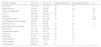
We analysed a total of 75 episodes in patients that visited the emergency department, of which 52% (39) were finally diagnosed as enterocolitis. Overall, diarrhoea was the most frequent presenting complaint (74.7%), followed by abdominal pain (56.4%), vomiting (54.7%), fever (53.3%), abdominal distension (48%) and lethargy or prostration (26.7%) (Table 2).
Clinical variables under study.
| Clinical variables | No, % (n) | Yes, % (n) | Enterocolitis (39), n | No enterocolitis (36), n | p |
|---|---|---|---|---|---|
| Diarrhoea | 25.3 (19) | 74.7 (56) | 29 | 27 | .5 |
| Abdominal pain | 44 (33) | 56 (42) | 22 | 20 | .47 |
| Abdominal distension | 52 (39) | 48 (36) | 27 | 9 | .25 |
| Fever | 46.6 (35) | 53.3 (40) | 24 | 16 | .4 |
| Vomiting | 45.3 (34) | 54.7 (41) | 21 | 20 | .48 |
| Lethargy/prostration | 73.3 (55) | 26.7 (20) | 18 | 2 | .000 |
| Final diagnosis of enterocolitis | 48 (36) | 52 (39) | |||
| Abdominal X-ray | 20 (15) | 80 (60) | 37 | 23 | |
| Abnormal X-rays out of total | 60 (36) | 40 (24) | 19 | 5 | .2 |
| Blood tests | 14.7 (11) | 85.3 (64) | 34 | 30 | |
| Positive stool culture | 90.7 (68) | 9.3 (7) | 0 | 7 | |
| Admission | 22.7 (17) | 77.3 (58) | 39 | 19 | |
| Bowel rest | 5.2 (3) | 94.8 (55) | 34 | 21 | |
| Antibiotherapy | 29.4 (17) | 70.6 (41) | 37 | 4 | |
| Urgent surgery | 98.7 (74) | 1.3 (1) | 1 | 0 | |
| Complications | 88 (66) | 12 (9) | 6 | 3 | .3 |
The two diagnostic tests performed most frequently were the complete blood count (85.3%) and plain abdominal radiograph (80%). The radiographic findings were abnormal in 24 of the 60 abdominal X-rays performed in these patients (40%). Of the 39 patients with a discharge diagnosis of enterocolitis, 20 had normal X-rays and 19 abnormal X-rays. The radiographic changes found in our patients included bowel loop dilation (32%), obstruction (5.3%) and ectopic air (1.3%). The results of stool culture were positive in 7 of the 75 episodes (9.3%), with 3 cases of infection by rotavirus, 2 of infection by Campylobacter, 1 of rotavirus and Aeromonas caviae coinfection, and 1 of infection by Cryptosporidium. Only 3 patients were examined by abdominal ultrasound, with unremarkable findings in all. Blood culture was performed in 12% of the episodes, with negative results in all.
Out of the 75 episodes, 58 (77.3%) led to admission to an inpatient ward. During hospitalisation, 94% of cases were managed with complete bowel rest and rectal irrigation and suction, and 70.6% with intravenous antibiotherapy. The documented complications were: intestinal perforation requiring urgent surgery in 1 patient, and acute severe to in 8 patients, 2 of who developed acute renal failure requiring admission in the intensive care unit.
When we analysed the association between variables, we found that the only one that was significantly associated with the diagnosis of enterocolitis was lethargy, found in 46.1% of patients with a discharge diagnosis of enterocolitis and only 5.6% of patients with other diagnoses (p<.01) (Table 2). When we performed univariate logistic regression to identify risk factors for an enterocolitis diagnosis, we found that the risk of enterocolitis was significantly increased in patients presenting with lethargy (OR, 14.57), abdominal distension (OR, 6.75) and abnormal findings in abdominal X-ray (OR, 5.83). These 3 factors continued to be significantly associated with enterocolitis when we fitted the 2 multivariate models mentioned above (Table 3).
Odds ratios obtained for the clinical variables under study.
| Univariate regression OR (95% CI) | p | Multivariate analysisa OR (95% CI) | ||||
|---|---|---|---|---|---|---|
| 1 | p | 2 | p | |||
| Lethargy/prostration | 14.57 (3.06–69.26) | .001 | 14.38 (2.43–84.84) | .003 | 17.28 (2.10–142.06) | .008 |
| Abdominal distension | 6.75 (2.44–18.64) | .000 | 7.97 (2.14–29.63) | .002 | 21.03 (3.36–131.32) | .001 |
| Abnormal X-ray | 5.83 (1.98–17.16) | .001 | 9.55 (2.35–38.77) | .002 | 12.02 (2.24–64.33) | .004 |
| Fever | 3.66 (0.85–15.65) | .080 | ||||
| Diarrhoea | 0.98 (0.17–5.63) | .988 | ||||
| Abdominal pain | 0.39 (0.07–2.03) | .26 | ||||
| Vomiting | 1.20 (0.26–5.43) | .809 | ||||
We fitted 2 regression models in the multivariate analysis:
(1) We only included variables with a p-value of less than 0.20 in the univariate analysis (as is customarily done in scientific research, and taking into account the small sample size).
(2) We included every variable that seemed relevant based on previous causal knowledge (vomiting, diarrhoea, fever, abdominal distension, lethargy, abdominal pain, abnormal radiographic findings).
Despite advances in its diagnosis and treatment, HAEC continues to be a significant cause of morbidity and mortality in patients with HD.3 It is important for clinicians to be aware of its epidemiology, warning signs and the approach to its diagnosis and management in the emergency setting for the purpose of early diagnosis and treatment, which is what makes this study relevant.11
Hirschsprung-associated enterocolitis occurs frequently in patients with HD. Some authors estimate that it affects up to 50% of these patients.4 This high incidence, combined with its potential and severe complications, may justify the significant rate of admission of patients with HD and suspected enterocolitis. In our series, 3 out of 4 patients with HD that sought care for gastrointestinal symptoms in the emergency department were admitted due to suspicion of HAEC. However, during the hospital stay, only 50% of those admitted received a diagnosis of HAEC.
There is published evidence suggesting that HAEC is more frequent in patients with long-segment aganglionosis.12–14 However, we found a balanced distribution in our sample: 52.4% of patients with HAEC had short-segment disease and 47.6% long-segment disease, as has also been found in other series.8 Some authors have proposed that the incidence of HAEC is lower in patients that have undergone surgical correction,15–17 but our data did not support this hypothesis, as 95% of the patients in our sample had undergone surgery and most of them experienced at least 1 episode of HAEC.
Although some authors have developed clinical scores with diagnostic criteria for HAEC,4 when it comes to the paediatric emergency care setting, as we mentioned above, it is more important to emphasise the need to maintain a high level of suspicion based on compatible symptoms, of which the most prevalent in our study was diarrhoea. It is also worth noting that lethargy was the symptom most frequently associated with a final diagnosis of HAEC in our series, which was consistent with the findings of other authors.4,15,18 We consider that patients with HD that present in the emergency department with diarrhoea and abdominal pain associated with neurologic symptoms should be admitted to hospital with a suspected diagnosis of HAEC.
When it comes to diagnostic tests, the most important technique in our series and in previous publications was plain abdominal radiography, whose use is recommended in emergency settings when there is clinical suspicion of HAEC.15 Dilated bowel loops, ectopic air or signs of bowel obstruction, while nonspecific, are considered highly indicative of HAEC and were significantly associated with the disease in our case series. We also found that in approximately half of cases of HAEC, the findings of plain abdominal radiography were normal, and therefore absence of radiographic abnormalities does not rule out enterocolitis. Other methods, such as abdominal ultrasound or computed tomography, have not proven cost-effective in the diagnosis of HAEC.19,20 Sepsis is the most severe complication of HAEC, and consequently guidelines recommend collection of a sample for blood culture. In our sample, blood culture was performed in few patients, although this finding may have been biased, as our study had a retrospective design and data are not always available for retrieval under these circumstances.
Based on the literature and the findings we have presented, we propose 2 possible clinical scenarios where HAEC should be suspected in the emergency setting: a first scenario where the clinical presentation is highly indicative of HAEC (patient with lethargy, explosive diarrhoea and abdominal pain and distension) in which, whatever the radiographic findings may be, admission and prompt initiation of medical treatment is recommended; and a second scenario, where the patient presents with less specific symptoms and normal radiographic findings, in which risk factors should be assessed and, when in doubt, the patient should be admitted for observation and re-evaluation in the next 24h.
There are some limitations to our study. Its design did not allow us to calculate incidence rates or estimate the frequency of risk factors, as the sample consisted solely of patients with HD that presented to our emergency department with compatible symptoms. Nevertheless, we believe that we obtained a representative and sizeable sample of patients with HD treated in our hospital, whose department of paediatric surgery is renowned for the treatment of this disease. Another limitation is the diagnosis of HAEC itself. Since stringent diagnostic criteria and clinical prediction rules were not used for its diagnosis, HAEC may have been overdiagnosed in the emergency department, leading to a higher than necessary rate of admission. Then again, we believe that the high morbidity and mortality associated with HAEC justify this approach as opposed to a more stringent one in the context of clinical practice in the emergency setting.
ConclusionsOne out of every 2 patients with HD that presented with gastrointestinal manifestations with or without fever in the emergency department received a final diagnosis of enterocolitis. Diarrhoea was the most frequent symptom, followed by abdominal pain and vomiting. The presence of lethargy, abdominal distension and/or abnormal findings in the abdominal X-ray support a diagnosis of HAEC.
Due to the severity of HAEC, maintaining a high degree of suspicion in the emergency setting is of utmost importance. Diagnostic tests should be performed in patients presenting with compatible symptoms, including an abdominal X-ray, although normal radiographic findings do not rule out HAEC. Treatment with bowel rest and antibiotherapy should be initiated early in patients whose clinical or radiographic features are clearly indicative of HAEC, while patients with less specific features should remain under observation while awaiting a final diagnosis.
Conflicts of interestThe authors have no conflicts of interest to declare.
Please cite this article as: Sellers M, Udaondo C, Moreno B, Martínez-Alés G, Díez J, Martínez L, et al. Enterocolitis asociada a enfermedad de Hirschsprung: estudio observacional sobre clínica y manejo en un servicio de urgencias hospitalarias. An Pediatr (Barc). 2018;88:329–334.
Previous presentation: this study was presented at the XXI Congress of the Sociedad Española de Urgencias Pediátricas; April 2016; Valencia, Spain.

Anales de Pediatría (English Edition) follows the Recommendations for the Conduct, Reporting, Editing and Publication of Scholarly Work in Medical Journals