


Turner syndrome (TS), which is associated with complete or partial monosomy of the X chromosome, is one of the most frequent and best-known chromosomal disorders. In addition to the typical features, such as short stature and amenorrhoea, affected girls are at higher risk of having other diseases compared to the general population, including autoimmune disorders. The association of TS with autoimmune thyroiditis, type 1 diabetes and inflammatory bowel disease has been solidly established, but its association with juvenile idiopathic arthritis (JIA) is not as well understood. We present the cases of 3 girls with TS and JIA managed in our hospital whose diagnosis elicited uncertainty or unnecessary treatments and the results of a literature review that confirms that while the association is infrequent, it is nevertheless well established.
The 3 patients had a 45 × 0 karyotype. In 2 cases, the diagnosis was made in the neonatal period, and in 1 case it was made antenatally by means of amniocentesis. All 3 patients started growth hormone replacement therapy at age 3–4 years. All 3 received the diagnosis of TS years before JIA was diagnosed, and the history of TS had not been considered significant in any of them for the purpose of referral to rheumatology.
A girl aged 7 years was referred for evaluation of monoarthritis in the left knee identified a month prior. In December 2016, the patient underwent arthrocentesis for obtention of a synovial fluid sample that had features consistent with inflammation (13 500 white blood cells [WBCs]/mm3; 80% mononuclear cells; glucose concentration, 49 mg/dL, protein concentration, 4.8 g/dL). The patient received a diagnosis of JIA and treatment with intraarticular corticosteroids. Three months later she had a relapse in the same knee, leading to initiation of treatment with subcutaneous methotrexate (MTX), with a good response. The girl remained in remission for the following 12 months, so MTX was discontinued. After 5 months without treatment, in January 2019 the patient experienced a relapse in the left knee, leading to resumption of treatment with MTX. She is currently in remission.
The second case corresponded to a girl that had undergone surgical correction of tetralogy of Fallot that was haemodynamically stable and presented at age 5 years with arthritis in the right knee concurrent with a streptococcal infection (scarlet fever). Two weeks later she presented with arthritis in the contralateral knee and was admitted to receive intravenous antibiotherapy. She did not improve with treatment, so she was referred to the department of rheumatology for evaluation. Given the history and course of disease, with persistence of mild synovitis in the right knee, the patient received a diagnosis of oligoarticular JIA. She underwent arthrocentesis in both knees, with examination of synovial fluid revealing inflammatory features (WBC count, 9500 cells/mm3; 80% polymorphonuclear cells; glucose, 52 mg/dL, protein concentration, 5.3 g/dL). The patient received intraarticular corticosteroids and exhibited a partial response, which prompted initiation of subcutaneous MTX. She is currently in remission.
The third patient was a girl aged 6 years with a personal history of monoarthritis in the right knee and a previous arthrocentesis that had revealed inflammatory features in the synovial fluid (WBC count, 8700 cells/mm3; 60% mononuclear cells; glucose concentration, 66 mg/dL, protein concentration, 5.2 g/dL). She was referred to the department of rheumatology and given a diagnosis of oligoarticular JIA in January 2019. She has been treated with intraarticular corticosteroid injections and is currently in remission without treatment.
All 3 patients had negative results of tests for detection of antinuclear antibodies (ANA), rheumatoid factor (RF) and HLA-B27 antigen. None had associated uveitis or other autoimmune disorders.
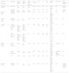
There are previous descriptions of the association between X monosomy and JIA in the literature (Table 1). Most articles on the subject correspond to single case reports,1–3 although there are also case series4,5 of up to 18 patients.6 The largest case series corresponds to a multicentre study with participation of 28 rheumatology centres in Europe and North America and followup of approximately 15 000 patients with JIA. Based on the number of identified patients, the authors estimated that the prevalence of the association of TS and JIA was six times greater than expected.6 A more recent study using the Danish Cytogenetic Central Register that reviewed a cohort of 798 women with TS followed up for 12,461 person‐years concluded that these patients had double the risk of developing an autoimmune disease compared to the general population, with a standardized incidence ratio for JIA (ratio of the observed number of cases to the expected number of cases) of 4.4 (Jørgensen et al., Arthritis Rheum. 2010;62:658–666).
Published cases of patients with Turner syndrome and juvenile idiopathic arthritis.
| Author (year of publication) | N | Karyotype | Age at diagnosis of TS | Age at diagnosis of JIA | Age at onset of joint symptoms | JIA oligo/poly | ANA | RF | HLA-B27 | Affected joints | Bone erosion | Other autoimmune diseases |
|---|---|---|---|---|---|---|---|---|---|---|---|---|
| Kohler et al (1981) | 2 | 46Xi (Xq) | 14 years | 15 years | 15 years | Oligo | NA | NA | Pos | Knees and 2 MCPs | NA | Crohn disease |
| 45X/46XX mosaicism | 15 years | 15 years | 15 years | Oligo | NA | NA | NA | Left knee | NA | Crohn disease | ||
| Balestrazzi et al (1986) | 1 | 45X/46XX mosaicism | 8 years | 8 years | 6 years | Oligo | 1/160 | NA | NA | Left knee., right 2nd finger, left 3rd finger. | NA | No |
| Foeldvari et al (1997) | 1 | 45X/46XX mosaicism | 10 years | 16 years | 8 years | Poly | NA | NA | Pos | Hands, wrists, elbows, knees and hips | No | No |
| Zulian et al (1998) | 18 | 11 Oligo, 45X (n = 6), 45X/46XX (n = 2), other (n = 3) | 1 month-17 years (mean 5 years and 5 months) | 1,7−12 years (mean 5 years and 8 months) | NA | Oligo | Pos 8/11 | Neg 8/8 | Neg in 5/5 | Knee (11/11), ankle (4/11), MCP/PIP (2/11), elbow (1/11) | No | NA |
| 7 Poly, 45X (n = 6) and 45X/46XX | 8 months-16 years (mean 6 years and 1 month) | 15 months-4.6 years (mean 3 years and 3 months) | NA | Poly | Pos 1/7 | Pos 1/7 | Neg in 1/1 | Knee 6/7, hip 6/7, ankle 5/7, wrist 3/7, cervical spine 1/7 | Yes | NA | ||
| Wilhborg et al (1999) | 3 | 45X/46X (Xq) mosaicism | 9 years | 12 years | 3 years | Poly | Neg | Neg | NA | Hands, wrists, knees, cervical and lumbar spine | Yes (feet) | No |
| 45X/46X (Xq) mosaicism | 6 years | 15 months | 15 months | Poly | Neg | Neg | NA | Hands, elbows, knees and feet | yes (carpals, ulna, elbows, metatarsals) | No | ||
| 45X | 1 month | 9 years | 9 years | Poly | Neg | Neg | NA | MCP, PIP, carpal bones, wrists, elbow, knees | Yes | No | ||
| Inamo et al (2000) | 1 | 45X/46XX mosaicism | NA | 14 years | 14 years | Poly | Neg | Neg | NA | bilateral PIPs, right elbow, wrists, right knee and ankle | NA | Diabetes mellitus, Hashimoto disease |
ANA, antinuclear antibodies; MCP, metacarpophalangeal joint; NA, data not available; Neg, negative; Oligo: oligoarticular; PIP, proximal interphalangeal joint; Poly: polyarticular; Pos, positive; RF, rheumatoid factor; TS, Turner syndrome.
The presentation of arthritis in JIA may be oligoarticular (up to 4 affected joints) or polyarticular (5 or more affected joints), with variability in the detection of ANA and HLA-B27 antigen and the presence of bone erosion. Additional autoimmune disorders are frequently reported, especially inflammatory bowel disease in HLA-B27-positive patients.
In conclusion, the prevalence of JIA in girls with TS is greater than expected for a random association. This must be taken into account in the evaluation of patients with TS that develop musculoskeletal manifestations suggestive of arthritis (joint swelling, limping or morning stiffness).
Please cite this article as: Lavilla P, Manzanares Á, Rabadán E, de Inocencio J. Artritis idiopática juvenil y síndromede Turner. An Pediatr (Barc). 2020;93:259–261.
