


A girl aged 8 years visited the Department of Otorhinolaryngology for assessment of snoring and obstructive sleep apnoea of 3 month’s duration. The examination of the oral cavity evinced bilateral tonsillar hypertrophy and a pedunculated mass originating in the lower pole of the right tonsil, with a smooth surface, pink hue and firm consistency. The patient had no cervical lymph node enlargement.
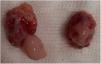
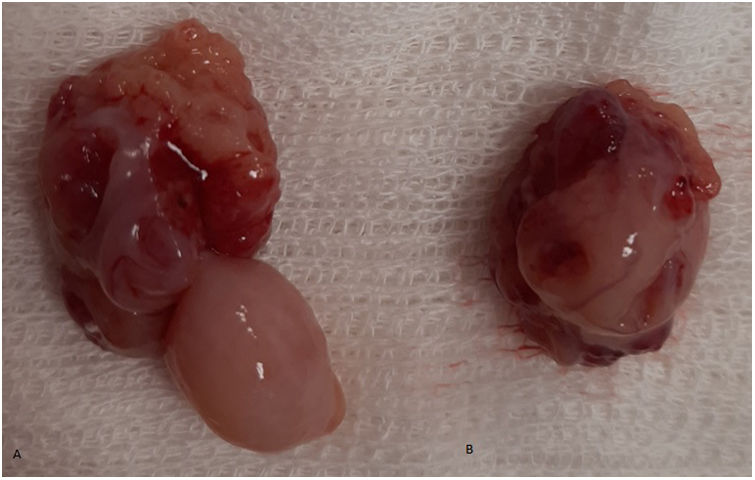
The patient underwent bilateral tonsillectomy under general anaesthesia, which included removal of the mass (Fig. 1).
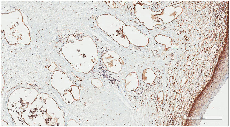
The histopathological examination identified a low-flow lymphatic malformation (lymphangioma) (International Society for the Study of Vascular Anomalies [ISSVA] classification)1 (Figs. 2 and 3).
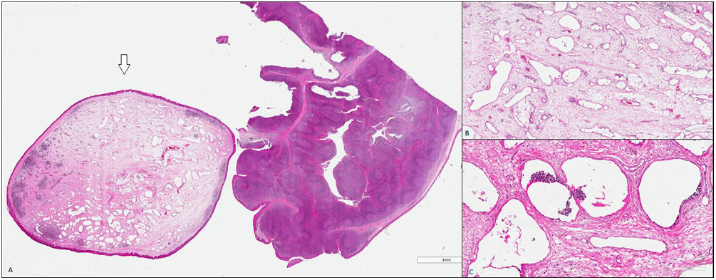
Microscopy. (A) Right tonsil with reactive lymphoid follicular hyperplasia and a polypoid outgrowth lined with squamous epithelium and, in the central portion, numerous vascular structures of a lymphatic nature with proteinaceous material and lymphocytes. No observable atypia. Haematoxylin and eosin (H&E) stain, original magnification ×10. (B and C) Lymphatic malformation. H&E, original magnification, ×20 (B) and ×40 (C).
There were no complications nor any evidence of the lesion recurring at 6 months of follow-up.
Lymphangiomas are congenital malformations rarely found in the tonsils, and account for 2% of all tonsillar masses.2 The differential diagnosis includes papilloma, lipoma and cyst, among others. The diagnosis is confirmed by the histopathological examination. They are usually unilateral and more frequent in male individuals.3 They may be asymptomatic or present with symptoms associated with local airway obstruction and irritation. They do not regress spontaneously and may become more apparent as the child grows.1 The usual treatment involves resection of the mass and tonsillectomy.2,3 There have been no reported cases of recurrence or malignant transformation.
