

In November 2014, an extended-spectrum beta-lactamase producing Klebsiella pneumoniae outbreak was detected in the neonatal intensive care unit of a tertiary care hospital.
ObjectiveOur aim was to determine the clinical, epidemiological and microbiological characteristics of the outbreak, to analyse the identified risk factors and to describe the preventive and control measures implemented for its eradication.
MethodsWe conducted a case-control study. We performed univariate and bivariate analyses, defining statistical significance as a P-value of less than 0.05. The implemented preventive and control measures were aimed at establishing the magnitude of the outbreak, effective communication, the evaluation of health care processes and education on patient safety. Clinical samples were collected for molecular and phenotypic characterisation.
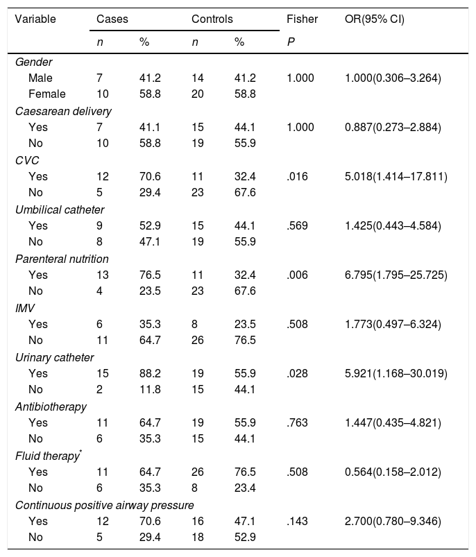
FindingsThe sample consisted of 51 newborns, of who 17 were cases and the remaining 34 controls. The distribution of cases by birth weight was: 2 cases (11.8%) greater than 2500g, 4 cases (23.5%) between 1500 and 2500g, 5 cases (29.4%) between 1000 and 1500g, and 5 cases (29.4%) less than 1000g. In one case, the birth weight was not documented in the health record. The following risk factors for colonisation or infection were statistically significant in our study: presence of a central venous catheter (OR, 5.0 [95% CI, 1.4–17.8]; P=.016); parenteral nutrition (OR, 6.8 [95% CI, 1.8–25.7]; P=.006); urinary catheterization (OR, 5.9 [95% CI, 1.2–30.0]; P=.028) and birth weight (P=.035). We found statistically significant differences in the mean total length of stay in hospital (P=.004) and length of stay in the NICU (P=.002). All 17 cases presented antimicrobial resistance with presence of extended-spectrum beta-lactamase type CTX-M-14.
ConclusionWorkplace interventions focused on patient safety need to be reinforced, especially those concerning practices with the potential to increase the extrinsic risk of colonisation or infection by extended-spectrum beta-lactamase-producing K. pneumoniae in the NICU, such as the insertion, care and maintenance of central venous catheter, parenteral nutrition and urinary catheterization.
En noviembre de 2014, se detectó un brote de Klebsiella pneumoniae productora de betalactamasas de espectro extendido en la unidad de cuidados intensivos neonatales de un hospital terciario.
ObjetivoEl objetivo fue describir las características clínicas, epidemiológicas y microbiológicas del brote, analizar los factores de riesgo asociados y presentar las medidas preventivas y de control implementadas para su erradicación.
MétodosEstudio de casos y controles. Se realizaron análisis univariantes y bivariantes, considerándose estadísticamente significativo un valour de p < 0,05. Las medidas preventivas y de control se centraron en la necesidad de determinar la magnitud del problema, en la comunicación efectiva, la evaluación de los procedimientos sanitarios y la educación sobre la seguridad del paciente. Se realizó caracterización molecular y fenotípica de muestras clínicas.
ResultadosLa muestra de estudio consistió en 51 neonatos, de los que 17 fueron casos y los 34 restantes controles. La distribución de casos por peso al nacer fue: 2 con peso > 2.500g (11,8%), 4 con peso de 1.500–2.500g (23,5%), 5 con peso de 1.000–1.500g (29,4%) y 5 con peso < 1.000g (29,4%). En un caso no se había registrado el peso al nacer en la historia clínica. Los factores de riesgo estadísticamente significativos para la colonización/infección fueron: presencia de catéter venoso (OR=5,0 [IC 95% 1,4–17,8]; p=0,016); nutrición parenteral (OR=6,8 [IC 95%: 1,8–25,7]; p=0,006); sondaje vesical (OR=5,9 [IC 95% 1,2–30,0]; p=0,028) y peso al nacer (p=0,035). Encontramos diferencias estadísticamente significativas en la estancia hospitalaria media (p=0,004) y los días de estancia en la unidad de cuidados intensivos neonatales (p=0,002). Se encontró resistencia a antimicrobianos tipo betalactamasas de espectro extendido CTX-M-14 en los 17 casos.
ConclusiónHan de reforzarse las iniciativas de trabajo sobre la seguridad de los pacientes, especialmente aquellas aplicables a intervenciones con el potencial de aumentar el riesgo de colonización/infección por K. pneumoniae productora de betalactamasas de espectro extendido en la unidad de cuidados intensivos neonatales, como las asociadas a la inserción, cuidado y mantenimiento de catéter venoso, la nutrición parenteral y el sondaje vesical.
The Enterobacteriaceae family constitutes an important and heterogeneous subgroup in the greater group of the gram-negative bacteria. They are facultative anaerobic microorganisms that live as saprophytes in the gastrointestinal tract, although they are also characterised by their ubiquity, as they are commonly found in the environment as well as in the GI tract of many animals.1
At present, Enterobacteriaceae are an important problem in many hospitals, as they are benefitting from several factors, such as the excessive use of antibiotics or prolonged hospital stays.2 There have been numerous reports of Klebsiella pneumoniae outbreaks in neonatal intensive care units (NICUs) in hospitals of different care levels throughout the world. This is indicative of the substantial complexity of preventing healthcare-associated infections (HAI) in these settings, which is compounded by the high morbidity and mortality associated with them.3
Articles on the subject published in recent years have identified several risk factors, described different strategies for outbreak control and reported morbidity and mortality rates with significant variations between studies. For instance, an outbreak that occurred in China in 2012 affected 103 newborns,4 an outbreak in India in 2010 had an associated mortality of 57%,5 and an outbreak in Germany in 2015 resulted in the permanent closure of the affected NICU.6 In addition, a systematic review published in 2016 by Stapleton et al. identified understaffing as the most important risk factor, and reported a median outbreak duration of 6.5 months.3
In November 2014, an extended-spectrum β-lactamase (ESBL)-producing Klebsiella pneumoniae outbreak was detected in the NICU of a tertiary hospital. The outbreak was terminated in January 2015 and involved a total of 17 cases (13 of colonisation and 4 of infection), none of which resulted in death. The control measures that were implemented achieved termination of the outbreak in less than 3 months.
The aim of this work is to analyse the clinical, epidemiological and microbiological characteristics of the outbreak and the associated risk factors, and to describe the measures of prevention and control that were implemented for its termination.
MethodologyStudy settingThe setting was a tertiary referral hospital located in the north of Spain. The Department of Neonatology operates under the Paediatrics Clinical Management Unit and is the reference unit for its geographical region. The department has a total of 32 beds, of which 14 belong to the NICU, and 2 are equipped for patient isolation. The usual occupancy rate is 50%.
Study designWe conducted an observational and analytical case–control study.
Case definitionWe established the following case definitions7:
- •
Infection by ESBL-producing K. pneumoniae: isolation of ESBL-producing K. pneumoniae from biological samples and features of infection that supported that this bacterium was the aetiological agent.
- •
Colonisation by ESBL-producing K. pneumoniae: isolation of ESBL-producing K. pneumoniae from biological samples in the absence of features of infection.
We selected two controls for each case. We used stratified sampling to maximise the homogeneity of cases and controls in relation to sex and birth weight. The period under study went from November 3, 2014 (when the first case was detected) to February 20, 2015 (when the last affected patient was discharged). Consequently, we selected the controls from the set of newborns admitted to the NICU in the same time interval.
We selected the controls from the patients admitted to the unit in the same time period as the cases using a random number generator known as nosetup.
Selection and definition of variablesWe performed a search of the PubMed database for articles on the subject of interest published in English in the past 10 years. We identified and reviewed 41 articles, and selected those variables found to be relevant in at least 25% of the articles for analysis. These variables were mainly related to placement of invasive devices.2,4,8–10 We also thought it would be of interest to include the variable ‘continuous positive airway pressure’ (CPAP) on account of its recent introduction in our NICUs.
Preventive and control measuresWith the aim of improving control of infection transmission and putting an end to the outbreak as quickly as possible, the Department of Preventive Medicine and Public Health developed a document compiling current recommendations for healthcare workers and relatives of patients.
- 1.
An epidemic curve was produced to identify the primary case and to track the epidemiological evolution of the outbreak (Fig. 1).
- 2.
Maps of the prevalence of multi-drug resistant microorganisms were drawn periodically to collect information on the evolution of the outbreak at specific time points. The maps covered both inpatient wards and NICU areas. They were shared with the medical and nursing management teams of the hospital and with physicians involved in any of the clinical departments of the hospital.
- 3.
Communication strategy for health care professionals and relatives of patients:
- a.
A document containing general recommendations for outbreak control was developed and given out to professionals involved in NICU operations and other workers. The document addressed aspects related to effective communication, the microbiological strategy, contact isolation and standard precautions, safety of health care procedures, health care clothing, the physical distribution of the space, routine work protocols, cleaning protocol, hands hygiene and management of patient visits.
- b.
The Department of Preventive Medicine and Public Health of the hospital held 2 information sessions for the families of the patients involved in the outbreak to explain the situation and the preventive measures that it was going to implement. The purpose of these sessions was to provide direct information about the interventions that were going to be implemented and to ask for their collaboration in minimising the transmission of these sessions was to present first-hand the actions that would be carried out and to achieve their collaboration in minimising pathogen transmission.
- a.
- 4.
Patients in the NICU were transferred to an alternative location to allow environmental decontamination of the unit with vaporised 35% hydrogen peroxide.

To determine the magnitude of the outbreak, pharyngeal and rectal samples were obtained from every patient that was staying in the NICU when the second case was detected. After that, samples were collected on a weekly basis from every patient whose previous results had been negative. Clinical samples were submitted to the National Centre of Microbiology for analysis. This laboratory characterised ESBL-producing K. pneumoniae isolates using phenotypic and molecular methods, including pulsed-field gel electrophoresis.
Samples of high-contact environmental surfaces, the hands of health care workers and disinfectants were obtained with sterile swabs and kept in liquid Amies medium. They were processed according to the recommendations of the Sociedad Española de Enfermedades Infecciosas y Microbiología Clínica in no 42 of its series of scientific documents on microbiological procedures, devoted to environmental microbiological control.11
Statistical analysisWe performed a descriptive analysis of each variable (univariate analysis), calculating the absolute and relative frequencies for qualitative variables. For quantitative variables, we obtained the mean as the measure of central tendency (with the corresponding standard deviation) as well as the minimum and maximum values.
We performed a bivariate analysis to assess potential associations in the variables under study. We used the Fisher exact test for dichotomous qualitative variables, as the sample was small and did not fulfil the necessary conditions for application of the chi square test. We used the non-parametric Mann–Whitney U test for quantitative variables, as the data did not follow a normal distribution based on the Shapiro–Wilk test (P<.001 in all four criteria and overall) and there were fewer than 30 individuals in the case group. We defined statistical significance as a P-value of less than .05.
We performed the analyses with the Statistical Package for the Social Sciences, version 18.0, and the software EPIDAT, version 3.1.
ResultsGeneral descriptionThe sample consisted of 51 newborns, out of who 17 corresponded to cases and the remaining 34 to controls. The sex distribution of the sample was 59% female and 41% male. The primary case, which was also the index case, was detected on November 22, 2014, and the last case on January 19, 2015. Of the 17 cases, 12 (70.5%) were of colonisation and 5 (29.4%) of infection (bacteraemia). Seven newborns in the case group (41.2%) had been delivered by caesarean section.
As for invasive medical procedures, umbilical venous catheters were used in 12 cases (70.6%), umbilical catheters in 9 (52.9%), parenteral nutrition (PN) in 13 (76.5%), invasive mechanical ventilation (IMV) in 6 (35.3%), continuous positive airway pressure (CPAP) in 12 (70.6%) and urinary catheterization (UC) in 15 (88.2%).
When it came to intravenous therapy, 11 patients in the case group (64.7%) received intravenous antibiotherapy, and 11 fluid therapy.
The distribution of patients with positive results for ESBL-producing K. pneumoniae was the following: 10 with a positive sample from a single location (58.8%), 5 with detection of the bacteria in samples from 2 locations (29.4%), and 2 with detection of the bacteria in samples from 3 or more locations (11.8%).
The distribution of cases by birth weight was: 2 (11.8%) with birth weights greater than 2500g, 4 (23.5%) with weights between 1500 and 2500g, 5 (29.4%) with weights between 1000 and 1500g and 5 (29.4%) with weights of less than 1000g. In 1 case, there was no record of the birth weight in the chart.
The distribution by gestational age was as follows: 6 (35.3%) full term, 2 (11.8%) early term, 8 (47.1%) late preterm and 1 (5.9%) extremely preterm.
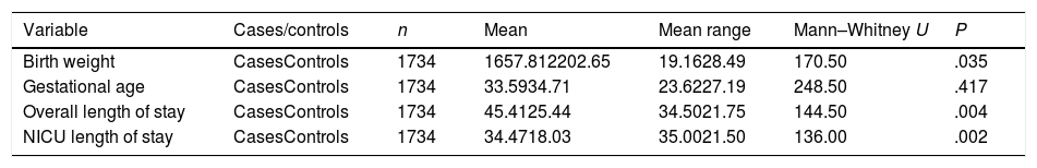
Last of all, the mean length of stay was 45.4 days (SD, ±23.6) for the overall stay in hospital and 34.5 days (SD, ±20.2) for the NICU stay.
Risk factorsThe risk factors with a statistically significant association with colonisation or infection by ESBL-producing K. pneumoniae were: CVC (OR, 5.0 [95% CI, 1.4–17.8]; P=.016], PN (OR, 6.8 [95% CI, 1.8–25.7]; P=.006); UC (OR, 5.9 [95% CI, 1.2–30.0]; P=.028) and birth weight (P=.035) (Table 1).
Risk factors associated with the presence of K. pneumoniae producer of ESBL.
| Variable | Cases | Controls | Fisher | OR(95% CI) | ||
|---|---|---|---|---|---|---|
| n | % | n | % | P | ||
| Gender | ||||||
| Male | 7 | 41.2 | 14 | 41.2 | 1.000 | 1.000(0.306–3.264) |
| Female | 10 | 58.8 | 20 | 58.8 | ||
| Caesarean delivery | ||||||
| Yes | 7 | 41.1 | 15 | 44.1 | 1.000 | 0.887(0.273–2.884) |
| No | 10 | 58.8 | 19 | 55.9 | ||
| CVC | ||||||
| Yes | 12 | 70.6 | 11 | 32.4 | .016 | 5.018(1.414–17.811) |
| No | 5 | 29.4 | 23 | 67.6 | ||
| Umbilical catheter | ||||||
| Yes | 9 | 52.9 | 15 | 44.1 | .569 | 1.425(0.443–4.584) |
| No | 8 | 47.1 | 19 | 55.9 | ||
| Parenteral nutrition | ||||||
| Yes | 13 | 76.5 | 11 | 32.4 | .006 | 6.795(1.795–25.725) |
| No | 4 | 23.5 | 23 | 67.6 | ||
| IMV | ||||||
| Yes | 6 | 35.3 | 8 | 23.5 | .508 | 1.773(0.497–6.324) |
| No | 11 | 64.7 | 26 | 76.5 | ||
| Urinary catheter | ||||||
| Yes | 15 | 88.2 | 19 | 55.9 | .028 | 5.921(1.168–30.019) |
| No | 2 | 11.8 | 15 | 44.1 | ||
| Antibiotherapy | ||||||
| Yes | 11 | 64.7 | 19 | 55.9 | .763 | 1.447(0.435–4.821) |
| No | 6 | 35.3 | 15 | 44.1 | ||
| Fluid therapy* | ||||||
| Yes | 11 | 64.7 | 26 | 76.5 | .508 | 0.564(0.158–2.012) |
| No | 6 | 35.3 | 8 | 23.4 | ||
| Continuous positive airway pressure | ||||||
| Yes | 12 | 70.6 | 16 | 47.1 | .143 | 2.700(0.780–9.346) |
| No | 5 | 29.4 | 18 | 52.9 | ||
CVC, central venous catheter; IMV, invasive mechanical ventilation.
We also found statistically significant differences in the mean overall length of stay and the mean length of stay in the NICU between cases and controls, with stays that were longer in the case group, by a mean of 20 days for the total hospitalisation (25 vs 45 days, P=.004) and a mean of 16 days for the stay at the NICU (18 vs 34 days, P=.002) (Table 2).
Mean differences between cases and controls.
| Variable | Cases/controls | n | Mean | Mean range | Mann–Whitney U | P |
|---|---|---|---|---|---|---|
| Birth weight | CasesControls | 1734 | 1657.812202.65 | 19.1628.49 | 170.50 | .035 |
| Gestational age | CasesControls | 1734 | 33.5934.71 | 23.6227.19 | 248.50 | .417 |
| Overall length of stay | CasesControls | 1734 | 45.4125.44 | 34.5021.75 | 144.50 | .004 |
| NICU length of stay | CasesControls | 1734 | 34.4718.03 | 35.0021.50 | 136.00 | .002 |
NICU, neonatal intensive care unit.
The main measures implemented were the following:
Information and communication management- •
A working group was appointed to manage the situation. It consisted of professionals from the NICU, the Department of Preventive Medicine and Public Health, the Department of Microbiology Department and the Cleaning Service.
- •
Communication strategy: immediately after the outbreak was detected, the situation was assessed in the course of several meetings that included the families of affected patients and professionals working in the involved units of the hospital. In these early meetings, families were informed about the characteristics of the outbreak, transmission mechanisms and the preventive measures that were going to be implemented, and they were asked to cooperate during the implementation phase. Meetings of parents, health care professionals and hospital management continued to be held regularly to share relevant information about the evolution of the outbreak and the measures taken to control it.
- •
Training plan and educational measures with participation of parents and other relatives of the patients.
- •
Collection of a total of 27 samples for microbiological testing: from work surfaces, hands of health care workers and cleaning and antiseptic products.
- •
Transfer of patients in the NICU to an alternative location: the infants were temporarily moved to a different location. Cases and controls were isolated separately. The NICU was disinfected though vaporisation of 35% hydrogen peroxide once the patients had been moved.
Testing found microbial resistance through production of ESBL type CTX-M-14 in all 17 cases. The molecular weight marker used in the pulsed-field gel electrophoresis tests led to the identification of 3 different profiles in the samples: 1 sample in group 1, 15 in group 2 and 1 in group 3.
All cultures of environmental samples were negative except for the cultures of a sample of topical gel and a sample taken from the hands of a health care worker, which grew the same microorganism involved in the outbreak: ESBL CTX-M-14-producing K. pneumoniae.
DiscussionIn this article, and in line with the initial objectives of our study, we have described a nosocomial outbreak that occurred in the NICU of a tertiary care hospital. We have analysed risk factors that may have been involved in the outbreak and detailed the measures implemented to control the outbreak in the shortest possible time.
The risk factors for infection or colonisation by ESBL-producing K. pneumoniae that we identified were birth weight and the use of any of 3 invasive procedures (CVC insertion, PN and UC). Our results were consistent with those of Maltezou et al.12 and González et al.,13 which found an association between low birth weight and infection by Klebsiella pneumoniae in outbreaks that developed in NICUs. However, other studies we reviewed had not found evidence of this association.6 It would make sense for low birth weight to be a risk factor for developing a HAI, as low birth weight newborns have lower T cell counts at birth and therefore could be more vulnerable to infection.14
The outbreak under study took place over a total of 57 days between November 22, 2014 and January 19, 2015. This is a relevant fact, since the final duration of the outbreak in our unit was considerably shorter compared to the findings of the only systematic review on infections by Enterobacteriaceae in NICU settings published in the literature, which reported a median outbreak duration of 6.2 months.3 We found several articles in our literature review that reported durations of even more than 6.2 months. In this sense, we ought to mention an outbreak that occurred in Greece in 2012, which lasted a total of 9 months,9 an outbreak in that occurred in New Guinea in 2007 with a duration of 13 months,15 and an outbreak in Italy in 2008 that lasted 26 months.16 In the last case, it should be taken into account that the outbreak affected 127 newborn infants, due to which it was extremely difficult to control.
We also ought to highlight an outbreak that occurred in Germany in 2015 that could not be controlled and resulted in the permanent closure of the affected NICU.6
On the other hand, a number of outbreaks of shorter or similar duration compared to the one in our unit have also been described, although the number of cases was lower in all of them.5,12,17,18 To measure the impact of the outbreak it is important to refer to mortality. None of the patients in our unit died, which differentiated this outbreak from most other outbreaks controlled within 2 months.5,12,17,18
Measures of prevention and control are the cornerstone of outbreak eradication. Therein lies the importance of strict adherence by all relevant parties and of effective communication between different work teams. It is also important to involve the families by keeping them updated and urging them to adhere to the recommended measures.19
The rapid termination of the outbreak was possible thanks to the urgency with which the Department of Preventive Medicine and Public Health developed adequate measures for control and, needless to say, to their correct implementation by health care professionals. Control was achieved through a health education plan implemented through the scheduling of multidisciplinary meetings whose purpose was to promote team work and emphasise the seriousness of the situation.
Health education and reinforcement of hand hygiene and other contact precautions were the interventions reported interventions in the reviewed literature.2,4–6,12,18,20
The reason is that they are effective and easily implemented interventions, and therefore their use for control of HAIs is a given. It is also of paramount importance to emphasise the need for hand hygiene routines using alcohol-based solutions, since currently this measure is only implemented in approximately 40% of the instances where it applies.21
On the other hand, organisational measures also played an important role, such as increasing the nurse-to-patient ratio, with some nurses allocated exclusively to the care of hospitalised patients affected by the outbreak, transfer of patients outside the NICU, screening of multi-drug resistant microorganisms in all newly-admitted patients, and physical isolation of patient cohorts.
It is also important to remember that family members that visit the NICU can also contribute to the spread of infection. Although the evidence on this aspect is mostly focused on respiratory infections caused by viruses,22 cases of transmission of Enterobacteriaceae through the hands of visitors have also been described in the literature.23 For this reason, we believe it may be beneficial to restrict access to the NICU, maintaining only the contact between the newborns and their parents.
Before concluding, we ought to note that there are limitations to our study. First of all, it was a case-control study, and therefore there is potential for information bias. Furthermore, due to the small sample size, the results of our study cannot be generalised.
In brief, invasive medical interventions such as insertion of CVCs, PN and UC together with low birth weight are risk factors for acquiring ESBL-producing K. pneumoniae, while colonisation or infection by ESBL-producing K. pneumoniae is associated with increases in the length of stay, with a mean increase of 20 days in overall hospital stay and 16 days in the NICU stay. Measures aimed at patient safety need to be reinforced in health care settings, with emphasis on those that address practices that may increase the extrinsic risk of colonisation or infection by ESBL-producing K. pneumoniae in NICUs.
Conflicts of interestThe authors have no conflicts of interest to declare.
Please cite this article as: Fernández-Prada M, Martínez-Ortega C, Santos-Simarro G, Morán-Álvarez P, Fernández-Verdugo A, Costa-Romero M. Brote de Klebsiella pneumoniae productora de betalactamasas de espectro extendido en una unidad de cuidados intensivos neonatales: factores de riesgo y medidas de prevención clave para su erradicación en tiempo récord. An Pediatr (Barc). 2019;91:13–20.
