


Magnetic resonance imaging (MRI) of the brain is a key tool in the diagnosis and prognosis of neurological conditions in newborn infants, especially when used along cranial ultrasound (cUS). Although its use has increased over the last decade, significant variations in protocols between centers persist. This document, developed by the Spanish Neonatal Brain Group in collaboration with pediatric neuroradiologists, establishes recommendations based on scientific evidence and clinical experience to standardize its use.
The document describes the logistics of neonatal MRI, including patient preparation, image acquisition, and condition-specific protocols. Clinical indications and optimal timing of MRI are outlined to maximize its diagnostic and prognostic value in different pathologies such as hypoxic-ischemic encephalopathy, metabolic encephalopathy, arterial infarction, venous sinus thrombosis, congenital infections and brain damage associated with hypoglycemia, hyperbilirubinemia or prematurity. Condition-specific patterns of damage and their prognostic correlation are described.
The document offers recommendations for the standardization of radiological reports and the reporting of critical findings to improve communication between radiologists, clinicians and parents.
In conclusion, these recommendations aim to optimize the use of brain MRI in neonatology, which will result in improved diagnostic accuracy and better-informed therapeutic decision-making, with the ultimate goal of improving neurodevelopmental outcomes in this vulnerable population.
La resonancia magnética (RM) cerebral es una herramienta clave en el diagnóstico y pronóstico de patologías neurológicas en recién nacidos, especialmente cuando se complementa con ecografía cerebral (EC). Aunque su uso ha aumentado en la última década, persisten variaciones significativas en los protocolos entre centros. Este documento, desarrollado por el Grupo Cerebro Neonatal Español en colaboración con neuroradiólogos pediátricos, establece recomendaciones basadas en evidencia científica y experiencia clínica para estandarizar su uso.
El documento describe la logística de la RM neonatal, incluyendo la preparación del paciente, adquisición de imágenes, y protocolos específicos según la patología. Se señalan las indicaciones clínicas y el momento óptimo para realizar la RM, con el fin de maximizar su valor diagnóstico y pronóstico en diferentes patologías como encefalopatía hipóxico-isquémica, metabólica, infarto cerebral arterial, trombosis del seno venoso, infecciones congénitas y daño neurológico asociado a hipoglucemia, hiperbilirrubinemia o prematuridad. Se describen los patrones de daño específicos de cada condición y su correlación pronóstica.
Asimismo, se proponen recomendaciones para la estandarización de informes radiológicos y la notificación de hallazgos críticos, con el fin de facilitar una mejor comunicación entre los radiólogos, los clínicos y las familias.
En conclusión, la implementación de esta propuesta busca optimizar el uso de la RM cerebral en neonatología, lo que se traducirá en diagnósticos más precisos y decisiones terapéuticas mejor informadas, con el objetivo final de mejorar los resultados neuroevolutivos en esta población vulnerable.
Magnetic resonance imaging (MRI), particularly when combined with cranial ultrasound (CUS), is the gold standard for brain imaging in the diagnosis and prognosis of neurologic disorders in neonates. Although no official data or survey results are available, the use of MRI in Spanish neonatal units has steadily increased over the past decade. However, there remains considerable heterogeneity among centers with respect to neuroimaging protocols, the timing of examinations, and the interpretation of findings. These aspects are fundamental, as they may determine the true clinical utility of the technique.
For these reasons, the Spanish Neonatal Brain Group-comprising professionals with particular expertise in neonatal neurologic disorders, in collaboration with pediatric neuroradiologists (Appendix B)-decided to develop a set of recommendations for the use of MRI in neonatal care. These recommendations are based on a synthesis of the available scientific evidence and the clinical and research experience of the group's members.
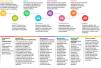
The objectives of this document are:
The objectives of this document are:
- •
To describe best practices and the minimum logistical requirements for centers to provide neonatal MRI, including patient preparation and image acquisition, in order to ensure that the procedure is safe for both neonates and accompanying healthcare staff.
- •
To provide recommendations on the clinical indications and optimal timing for neonatal brain MRI.
- •
To promote a standardized approach to reporting MRI findings, helping clinicians to assess their severity, anticipate their impact on neurodevelopmental outcomes, and adequately inform families.
- •
To achieve these objectives, eight working groups were created. Each one reviewed the literature relevant to a specific neonatal neurologic condition and summarized the available evidence. The full group then established the corresponding recommendations by consensus.
In most neonatal neurologic disorders, cranial CUS and MRI are complementary. CUS is the first-line imaging modality because of its bedside availability and its capacity for early and serial assessments.
The role of computed tomography (CT) in neonatal neurology is very limited. Except in surgical emergencies where MRI is not readily accessible, CT should not be used in neonates.1 It offers no diagnostic or prognostic advantage over the combination of MRI and CUS, and the risks from ionizing radiation are considerable and increasingly well documented. A recent European multicenter study reported a positive linear dose-response relationship between childhood head irradiation and the risk of malignant brain tumors, particularly glioblastomas.2
Requesting a brain MRITo ensure optimal assessment and interpretation of neuroimaging findings, MRI requests submitted to the neuroradiology team should include key clinical information about the patient: gestational age at birth, current postmenstrual and chronological age; relevant events since birth and their timing; any significant comorbidities; neurological signs, if present; and head circumference (including the corresponding percentile). It is also advisable to indicate any suspected clinical condition or specific lesion that needs to be ruled out, as this helps guide the selection of the most appropriate MRI protocol. Direct communication with the radiology team can further support this process.
Patient preparation for MRI and care during and after the ProcedureBrain MRI without sedation is a safe procedure in neonates and, when performed appropriately, can produce high-quality images. However, this requires a well-defined imaging protocol and adequate training of the healthcare team. Whenever possible, this should be the preferred modality.
Table S1 in Appendix B outlines the nursing care required before, during, and after the MRI examination.
Image AcquisitionHigh-quality imaging is essential for obtaining accurate and clinically useful information. At a minimum, this requires a 1.5 T MRI scanner, along with a dedicated neonatal head coil. If this is not available, a multichannel head coil that allows proper positioning and centering of the neonate should be used.
The imaging protocol—including the choice of sequences and their parameters—must be optimized for the neonatal brain. It should enable the detection of a wide range of conditions and be tailored to the patient's clinical presentation and history. Although a basic neonatal protocol is shown in Table 1, additional sequences should be included as needed on an individual basis (Table 2).
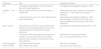
Basic neonatal brain MRI protocol.
| Sequence | Utility | Suggested parameters |
|---|---|---|
| Axial and coronal T2-weighted spin echo (T2SE) sequences | Assessment of brain anatomy and white matter maturation. It allows assessment of the cortex and detection of migration disorders. Also allows visualization of hypoxic-ischemic and hemorrhagic lesions. | TR 4300 |
| TE 135 | ||
| Axial diffusion weighted imaging (with ADC mapping) | Allows detection of recent acute hypoxic-ischemic and inflammatory lesions. | B 1000 |
| Axial and sagittal T1-weighted gradient echo images (may be obtained from 3D T1-weighted MRI) | Assessment of brain anatomy and myelination. Allows visualization of hypoxic-ischemic and hemorrhagic lesions. | TR 9.9 |
| TE 4.2 | ||
| Axial T2*-weighted gradient-recalled echo sequence or susceptibility-weighted sequence | Allows visualization of hemorrhagic lesions, calcifications and vascular anomalies. |
Abbreviations: ADC, apparent diffusion coefficient; TE, time to echo; TR, time to repetition.
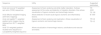
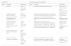
Indications for neonatal brain MRI.
| Condition | Indications | Optimal timing of imaging | Special sequences (in addition to basic protocol [Table 1]) |
|---|---|---|---|
| Acute encephalopathy (other than HIE) or seizures of unknown etiology | All patients | As soon as possible | MRS in all cases |
| Hypoxic-ischemic encephalopathy | All patients treated with TH Neonates with mild HIE with an atypical presentation or abnormal CUS findings | Optimal: 4−7 days post birth Valid: 4−14 days post birth Early (2−4 days) if redirection of care is considered | Consider MRS, voxel-based mapping of BGT T2-axial: 2 mm-thick slices needed for visualization of mammillary bodies |
| Neonatal ischemic stroke | Clinical presentation (clonic seizure or apnea) Suggestive sonographic findings (increased echogenicity corresponding to a vascular territory) | Within a week of onset | Cerebral and cervical non-contrast MR angiography (3D-TOF) to assess for carotid artery dissection, occlusion or stenosis of large or medium vessels or vascular anomalies SWI allows detection of clots |
| Cerebral venous thrombosis | Clinical presentation Suggestive sonographic findings | Within a week of onset | SWI allows visualization of congested small cerebral veins and isolated congestion or thrombosis of deep medullary veins and superficial cerebral veins Phase-contrast MR venography (if CVST is suspected) |
| Cerebral venous sinus thrombosis | Clinical presentation Suggestive sonographic findings (especially intraventricular hemorrhage, thalamic hemorrhage or white matter involvement) | As soon as possible after suspecting the condition if treatment with anticoagulants is considered. No anticoagulation: 5−7 days later to assess progression and delineate associated parenchymal lesions. If anticoagulation: consider monitoring to discontinue treatment at 6 weeks and, if thrombus persists, at 12 weeks | SWI allows visualization of intraluminal clot; significant asymmetries in vascular perfusion Venogram: assessment of venous flow |
| Meningoencephalitis | Abnormal or suggestive findings on CUS Complicated course | Depending on clinical and sonographic features If viral infection is suspected: first week (repeating 2−3 weeks later if initial results were abnormal) | |
| Congenital heart disease | Neurologic manifestations Abnormal or suggestive findings in CUS. Consider after extracorporeal surgery | As soon as the patient’s condition allows | |
| Congenital cytomegalovirus infection | Infection acquired during the first trimester of pregnancy Symptomatic infection (including neonates with isolated CNS infection) Neonates with abnormal CUS findings (lenticulostriate vasculopathy, ventriculomegaly, intraventricular septations, subependymal, periventricular or temporal cysts, abnormal white matter echogenicity, corpus callosum dysgenesis) | Urgent if there are doubts whether or not to start treatment As soon as possible in all other cases | SWI and GRE: make it easier to visualize calcifications |
| Neurometabolic disease | In every case | As soon as possible after it is suspected | MRS: single-voxel spectroscopy with a short TE can detect metabolites at low concentrations. Place the voxel in acute lesions with restricted restriction and in the BGT and, optionally, the parietal WM or mid-parietal gray matter. Avoid areas with chronic lesions secondary necrosis, hemorrhage or calcifications. |
| Hypoglycemia | Neurologic dysfunction (encephalopathy, seizures) Sonographic findings suggestive of injury | Within 2 weeks | |
| Hyperbilirubinemia | Acute neurologic manifestations Consider on a case-to-case basis if persistent severe hyperbilirubinemia requiring exchange transfusion or abnormal evoked potentials | At term-equivalent age Repeat at around 6−9 months in case of normal or uncertain neonatal MRI and abnormal psychomotor development | |
| Suspected CNS structural anomaly | Neurologic manifestations, craniofacial features Suggestive sonographic findings | Whenever possible Consider repeating later if there is uncertainty, especially in case of suspected cortical development abnormalities or in preterm infants. | Consider including non-contrast enhanced MR angiography of the brain |
| Preterm brain injury (see Table 7) | (1) Neurologic manifestations (especially cramped-snchronized general movements or other motor or behavioral warning signs at term-equivalent age), or (2) Evidence in CUS performed at term-equivalent age of WM injury (persistent or cystic abnormality in WM), stroke, posthemorrhagic ventricular dilatation, cerebellar hypoplasia/hemorrhage In these cases, brain MRI may help determine the nature and extent of injury better and do a more accurate prognosis. | At term-equivalent age, around 40 weeks postmenstrual age Before term-equivalent age if redirection of care is considered |
Abbreviations: BGT, basal ganglia and thalamus; CNS, central nervous system; CUS, cranial ultrasound; CVST, cerebral venous sinus thrombosis; GRE, gradient-recalled echo sequence; HIE, hypoxic-ischemic encephalopathy; MRS, magnetic resonance spectroscopy; PT, preterm; TE, time to echo; TH, therapeutic hypothermia; TOF, time of flight; SWI, susceptibility-weighted imaging; WM, white matter.
This section, along with Tables 2 to 7 and Table S2 in the supplementary material, describes the neonatal neurological conditions in which MRI plays a key role in diagnosis and prognosis.
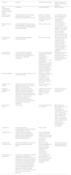
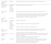
Magnetic resonance imaging findings in HIE and correlation with neurodevelopmental outcomes.
| Sequences | Anatomical region | Assessment | Prognostic correlation |
|---|---|---|---|
| Conventional and diffusion MRI Qualitative and systematic assessment of the following structures | Basal ganglia and thalamus | Assess for potential injury and its extension | The presence and severity of injury is associated with the risk and severity of motor impairment |
| Posterior limb of internal capsule | Assess for appropriate myelination for gestational age. | Abnormal myelination is a highly sensitive risk factor for motor impairment | |
| Brainstem | Assess involvement and extent of injury | Increases the severity of motor problems and is associated with an increased mortality | |
| White matter and cortex | Assess for abnormalities in signal intensity, whether they are focal or diffuse, and their extent | White matter injury increases the risk of visual, cognitive and behavioral problems. When it is very extensive, it is associated with mild CP. | |
| Mammillary bodies | Assess whether there is an increase in signal intensity in the axial or coronal T2-weighted images (requires 2 mm slice thickness). | Injury in the mammillary bodies increases the risk of learning and memory problems in school-aged children, even when the rest of the structures appear normal in the MRI | |
| Spectroscopy Single voxel Short TE (35 ms) | Basal ganglia-thalamus | Lactate-threonine/NAA levels correlate to neurodevelopmental outcomes at age 2 years | Values equal to or greater than 0.39 for lactate/NAA have been found to offer: - Sen 100% and Spe 97% for predicting motor impairment - Sen 90% and Spe 97% for predicting cognitive impairment - Sen 81% and Spe 97% for predicting language disorder |
Abbreviations: CP, cerebral palsy; HIE, hypoxic-ischemic encephalopathy; MR, magnetic resonance; NAA, N-acetyl-aspartate; Sen, sensitivity; Spe, specificity; TE, time to echo.
MRI findings in neonatal arterial ischemic stroke and associated prognosis.
| Sequences | Sign | Prognostic correlation |
|---|---|---|
| DWI | Pre-Wallerian degeneration in the corticospinal tract and cerebral peduncles | Contralateral monoplegia/hemiplegia in > 80% |
| [1,0]DWI, T1 and T2 | Massive stroke including ventricular collapse or midline shift | Moderate-severe motor, cognitive, and language impairment in > 70% High risk of epilepsy in childhood |
| Involvement of the main trunk of the middle cerebral artery and thalamus | Contralateral monoplegia/hemiplegia in > 90% Risk of epilepsy in childhood: 40% High risk of other neurodevelopmental disorders and visual impairment | |
| [2,0]T1 and T2 | Pyramidal tract involvement: lesion anterior to the central sulcus, basal ganglia, posterior limb of the internal capsule, or involvement of cerebral peduncles | Contralateral monoplegia/hemiplegia in 80 %–100 % |
| Involvement of the supramarginal and angular gyri | Increased risk of language disorders | |
| Infarction of the posterior portion of the middle cerebral artery or posterior cerebral artery; or the optic radiations | Risk of visual impairment (30 %–50 %) | |
| [1,0]Tractography at age 3 months | Corticospinal tract asymmetry | High risk of hemiplegia |
| Optic radiation asymmetry | High risk of visual impairment |
Abbreviations: DWI, diffusion-weighted imaging; MRI, magnetic resonance imaging; NAIS, neonatal arterial ischemic stroke.
Main neuroimaging features in congenital and neonatal viral infections.
| Infection | Main neuroimaging features |
|---|---|
| Zika virus | Microcephaly, calcifications (predominantly at the junction of the cortex and subcortical white matter), ventriculomegaly |
| Toxoplasmosis | Hydrocephalus (obstruction of cerebral aqueduct secondary to elevated CSF protein concentration and ependymitis). Diffuse nodular calcifications (secondary to granulomatous necrosis). Periventricular, cortical/subcortical or basal ganglia/thalamus involvement. |
| Herpes simplex virus | Encephalitis (characterized by marked edema and rapid progression to multicystic encephalomalacia). In neonates there is no predilection for the temporal lobes, as the infection is typically via hematogenous spread rather than by retrograde axonal transport from the oropharyngeal mucosa. |
| Parvovirus B19 | Ischemic or hemorrhagic complications associated with anemia or fetal transfusions. Reported cases of neurodevelopmental disorders and cortical malformation. |
| Rubella | Necrotizing vasculopathy. White matter injury (typically involving the temporal lobes). |
| Varicella zoster | Necrotizing encephalitis. Calcifications. |
| Treponema pallidum | Chronic meningovascular neurosyphilis |
| Lymphocytic choriomeningitis | Periventricular calcifications. Ventriculomegaly. |
| Enterovirus and parechovirus | Neurotropic viruses that typically cause white matter changes. The differential diagnosis with hypoxic-ischemic encephalopathy may be challenging. |
| Rotavirus | Damage caused by remote induction of interleukin-6 production in the CNS without direct infection of it. Similar pattern to those of enterovirus or parechovirus infection. Severe cases may progress to leukomalacia. |
| SARS-CoV-2 | Transplacental transmission is rare but possible, especially in the last weeks of pregnancy. There are a few isolated reports of transplacental transmission with neurologic involvement (white matter hyperintensities on MRI). |
CNS, central nervous system.
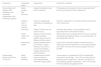
Indications and interpretation of neonatal brain MRI in preterm infants.
| Clinical scenario / sonographic finding | Indications | Important aspects that should be assessed and included in the radiology report |
|---|---|---|
| No evidence of injury on cranial ultrasound | Complicated neonatal coursea | Assess brain maturation with specific scales 36 |
| Abnormal neurologic examination | ||
| Noncystic WM injury | In all cases | Determine the severity of white matter injury using specific scoring systems 34,36,37 |
| If not possible, report the number of lesions, type (punctate or cystic), location, myelination and symmetry of the posterior limb of the internal capsule, ventricular morphology and size, corpus callosum size, biparietal diameter, and interhemispheric distance. | ||
| In addition, assess brain maturation using specific scales. | ||
| Cystic WM lesion | In all cases | |
| Germinal matrix hemorrhage/HIV I-II | Complicated neonatal course | Grade hemorrhage severity |
| Abnormal neurologic examination | Assess for and describe any other potential parenchymal lesions | |
| Other risk factors | In addition, assess brain maturation using specific scales. | |
| HIV III/periventricular infarction | In all cases | Report the location, extent (number of affected territories), midline shift, unilateral or bilateral involvement, motor pathway involvement and level of involvement. |
| In addition, assess brain maturation using specific scales. | ||
| Posthemorrhagic ventricular dilatation | In all cases | Description of ventricular size and morphology compared to previous studies. In patients with ventriculoperitoneal shunts or ventricular access devices, specify the location and position of the catheter tip. |
| Assess for and describe any other potential parenchymal lesions | ||
| In addition, assess brain maturation using specific scales. | ||
| Cerebellar injury | In all cases | Presence or absence of hemorrhages, size and side (left/right/bilateral) of involvement, measurement of transcerebellar diameter, presence or absence of signs of atrophy |
| Assess for and describe any other potential parenchymal lesions | ||
| In addition, assess brain maturation using specific scales. |
These clinical scenarios are derived from the cranial ultrasound screening program that should be applied to all preterm neonates admitted to neonatal units.
Abbreviations: HIV, intraventricular hemorrhage; MRI, magnetic resonance imaging; WM, white matter.
a Neonatal course is considered complicated, and therefore associated with increased neurologic risk, when three or more of the following factors are present: treated retinopathy, moderate-to-severe bronchopulmonary dysplasia (or supplemental oxygen requirement at 28 days after birth), necrotizing enterocolitis, sepsis, need for vasoactive drugs or corticosteroids, or hyperbilirubinemia requiring exchange transfusion.
Any preterm neonate presenting with a specific neurologic condition (e.g., hypoxic-ischemic encephalopathy, arterial infarction, meningitis) should be evaluated according to the protocol for full-term infants, as specified in Table 2, although neuroimaging findings must always be interpreted in relation to gestational age.
The presence of three or more of the following risk factors would be considered a complicated neonatal course for the purposes of increased neurologic risk: retinopathy requiring surgery, moderate-to-severe bronchopulmonary dysplasia (or need for supplemental oxygen at 28 days post birth), necrotizing enterocolitis, sepsis, need for vasoactive drugs or corticosteroids and hyperbilirubinemia in the exchange transfusion range.
It also addresses two common neurological emergencies in which early diagnosis is essential: acute neonatal encephalopathy and neonatal seizures.
Acute neonatal encephalopathy is a clinical syndrome characterized by altered alertness or neurobehavior, which may be accompanied by abnormal tone or reflexes, abnormal movement patterns, and, in some cases, seizures. It is a descriptive umbrella term that indicates neurological dysfunction of the brain, without implying a single cause. The underlying etiology may be hypoxic-ischemic, infectious, genetic, or metabolic.
In any neonate with acute neonatal encephalopathy or seizures where a hypoxic-ischemic cause is not suspected, MRI should be performed as early as possible. The goal is to rapidly guide diagnosis and, in some cases, treatment. This initial scan does not preclude the need for repeat imaging later on, if necessary.
Hypoxic-Ischemic EncephalopathyMRI is the gold standard for assessing the nature and severity of brain injury in hypoxic-ischemic encephalopathy (HIE), as well as for providing prognostic information.3
Hypoxic-ischemic injury is associated with characteristic lesion patterns (topographic distribution) that vary depending on gestational age, as well as the nature, severity, and timing of the insult.3,4 These injury patterns are strongly correlated with neurodevelopmental outcomes,4,5 making their accurate description in the MRI report essential (Table 3). Several MRI-based scoring systems with prognostic value? have been developed, primarily for research purposes, and require specific training. Although they can be useful, their use is not essential as long as the involvement of individual brain structures is described in sufficient detail.
In addition to the typical involvement of the cortex, basal ganglia, white matter, and brainstem, acute injury to the mammillary bodies has recently been described in the context of HIE. These lesions may be present alongside other brain injuries, but they have also been observed in neonates with otherwise normal imaging findings. Identifying and reporting these lesions is important, as they are associated with learning and memory difficulties at school age. Adequate visualization requires axial or coronal T2-weighted images with 2 mm slice thickness (Tables 2 and 3).4,6
Although CUS is routinely used in neonates with HIE, its sensitivity is limited--particularly for detecting cortical, cerebellar, and brainstem lesions–and its prognostic utility is also more restricted. Nonetheless, CUS is a complementary modality to MRI, allowing for early and serial evaluations and, thus, monitoring of brain injury over time. Spectral Doppler analysis of flow velocity waveforms can help detect patterns indicative of impaired cerebral autoregulation. A validated CUS scoring system is available and has been associated with neurodevelopmental outcomes in neonates with HIE.7
Neonatal strokeBrain MRI is the imaging modality of choice for establishing both the diagnosis of neonatal arterial ischemic stroke (NAIS)8 and its prognosis, based on: (1) the arterial territory predominantly affected, (2) the extent of the infarction, and (3) the anatomical or functional structures involved (Table 4).9–12
Until MRI can be performed, CUS may be used to detect NAIS, particularly middle cerebral artery infarction, provided it is performed ?24 hours after the event by an experienced operator with appropriate equipment.13
In cases where the neonatal MRI is inconclusive or when unexpected motor development is observed, repeat brain MRI with diffusion tensor imaging -based tractography at around 3 months of age can provide a more accurate prediction of neurodevelopmental outcomes14 (Table 4).
Cerebral venous infarction and venous sinus thrombosisConventional MRI combined with venography has a sensitivity of nearly 100% for detecting cerebral venous sinus thrombosis (CVST) in pediatric patients, although sensitivity is somewhat lower in neonates.15 MRI with venography should be performed urgently when CVST is suspected, as it also facilitates characterization of associated lesions. CUS has shown high specificity for the diagnosis of CVST16 and helps reduce false positives in MRI assessment.
MRI and CUS are complementary modalities for both diagnosis and follow-up, particularly for monitoring vessel recanalization.
Cerebral venous infarction most often results from obstruction of a cerebral vein and, less commonly, from hemorrhagic transformation of a neonatal arterial ischemic stroke. Venous infarctions are readily distinguished by their hemorrhagic nature and by their location within regions drained by deep medullary veins.
MRI studies are essential for diagnosis, with susceptibility-weighted imaging (SWI) playing a particularly important role. The prognosis of cerebral venous infarction depends on its location and on the structures involved within the lesion area.
Neonates with congenital heart diseaseBrain dysmaturation (secondary to sustained abnormalities in cerebral blood flow and metabolism during gestation), cumulative ischemia-reperfusion injury and acquired brain damage (eg, ischemic or hemorrhagic stroke) are the main pathogenic mechanisms involved in neurodevelopmental disorders detected in infants with congenital heart disease.17–19
While all neonates with congenital heart disease should be monitored with frequent CUS examinations, CUS must be specifically performed at the following time points: (1) at birth, to assess brain maturation and for the presence of prenatal lesions; (2) after each surgery or interventional procedure, to rule out complications; (3) if seizures or other neurologic manifestations develop; and (4) if there is acute hemodynamic deterioration. Whenever the sonographic findings are abnormal or inconclusive, the evaluation should be completed with MRI.
Congenital cytomegalovirus infectionCranial ultrasound and MRI are complementary tools in the study of congenital cytomegalovirus infection of the central nervous system (CNS).20,21 Cranial ultrasound is superior to MRI for detection of calcifications and lenticulostriate vasculopathy. All neonates with cytomegalovirus infection who are symptomatic or with abnormal CUS findings should undergo brain MRI to assess the extension of WM injury and rule out cortical or cerebellar malformations.22
Although there are no studies in which antiviral treatment was assigned based on the severity of the imaging findings, scores of 2 or 3 on neuroimaging severity scales generally constitute an indication for treatment (Table 5). For detailed information on the antiviral treatment protocol, we refer the reader to the applicable guidelines.25
Fetal and neonatal neuroimaging scoring systems for congenital CMV infection.
| Fetal MRI 23 | Postnatal ultrasound and MRI22,24 | ||||
|---|---|---|---|---|---|
| Score | Findings | Outcomes | Score | Findings | Outcomes |
| 1 | Normal | SNHL: 0 Neurologic sequelae: 1.6% | 0 | None of the following abnormalities | Normal: 87.5% Mild disability: 6.3% Moderate/severe disability: 6.3% |
| 2 | Isolated increased signal intensity of periventricular frontal or parietooccipital white matter on T2 | SNHL: 0 Neurologic sequelae: 0 | 1 | Single punctate periventricular calcification, lenticulostriate vasculopathy, caudothalamic germinolysis, mild ventriculomegaly or multifocal white matter signal abnormality on MRI | Normal: 83.7% Mild disability: 8.2% Moderate/severe disability: 8.2% |
| 3 | Isolated increased signal intensity of periventricular temporal white matter on T2 | SNHL: 14.3% Neurologic sequelae: 0 | 2 | Multiple discrete periventricular calcifications, paraventricular germinolytic cysts, occipital horn septations, significant ventriculomegaly, diffuse white matter signal abnormality or temporal lobe involvement | Normal: 48.7% Mild disability: 17.9% Moderate/severe disability: 33.3% |
| 4 | Cysts or septations in temporal or occipital lobe | SNHL: 55% Neurologic sequelae: 25% | |||
| 5 | Migration disorders, cerebellar hypoplasia | SNHL: 66.7% Neurologic sequelae: 66.7% | 3 | Extensive calcifications, brain atrophy, cortical malformation, dysgenesis of the corpus callosum or cerebellar hypoplasia | Normal: 3% Mild disability: 0 Moderate/severe disability: 97% |
Abbreviations: cCMV, congenital cytomegalovirus infection; MRI: magnetic resonance imaging; SNHL, sensorineural hearing loss.
Table 6 summarizes the neuroimaging findings associated with other CNS infections.
Neonatal meningoencephalitisAssessment with MRI is indicated in newborns with complicated meningitis or abnormal findings on the CUS. The optimal timing for MRI depends on the clinical condition of the patient and the changes in sonographic features over time. Magnetic resonance imaging is more reliable for diagnosis of extra-axial bacterial empyema.26,27
There are limited data on the correlation of neuroimaging findings with subsequent neurological outcomes, so the prognosis must be individualized and made with caution, taking into account all the clinical information for the patient, as opposed to just the imaging data.
Meningoencephalitis secondary to enterovirus, parechovirus, herpesvirus or rotavirus infection typically involves the WM (Table 6). In the case of abnormal WM echogenicity on the CUS or presence of neurologic manifestations, diffusion-weighted imaging would be indicated within a week of onset to assess for the characteristic pattern of these infections in the neonatal period: restricted diffusion typically involving periventricular and subcortical WM (and frequently frontal WM), corpus callosum, internal and external capsule, pyramidal tracts and cerebral peduncles, sparing the basal ganglia and cerebellum. Punctate ischemic lesions are also frequently detected (Table 6).
The evolution of abnormal imaging findings varies, with a tendency toward normalization in some cases and progression to multicystic encephalomalacia and gliosis in others. For this reason, acute-phase neuroimaging findings offer little prognostic value, and repetition of MRI 2–3 weeks later is recommended to assess for the presence of residual WM lesions.
Brain injury due to hypoglycemiaSevere, sustained, or repeated hypoglycemia can cause brain injury, especially involving the WM (including parenchymal hemorrhage) and cortex, but also the thalamus and globus pallidus.28 The blood glucose levels and episode durations associated with brain injury vary from patient to patient. Although it is not always the case, glucose levels that are low enough to cause brain injury are frequently accompanied by acute neurologic impairment and, in some cases, seizures.
Cranial ultrasound can detect changes in WM or basal nuclei echogenicity suggestive of injury. This requires confirmation by MRI. Normal CUS findings (especially if the assessment did not include images acquired through the posterior fontanelle) do not rule out occipital or posterior parietal injury. Therefore, in cases of severe and sustained hypoglycemia or in the presence of neurologic, MRI should be performed, even in patients with normal CUS findings.28
Brain injury secondary to hyperbilirubinemiaThe serum bilirubin levels that can cause brain injury–most commonly in the globus pallidus and subthalamic nucleus, though periventricular white matter involvement has also been reported29–depend on gestational age, postnatal age, and the presence of comorbidities. Most neonates who later develop neurodevelopmental disorders have a history of acute encephalopathy during the neonatal period, although in preterm infants the signs are subtler and more difficult to recognize.
CUS has low sensitivity for detecting basal ganglia injury, and normal CUS findings do not rule it out. Brain MRI should be performed in the neonatal period in all neonates with a history of encephalopathy, and considered on an individual basis in those who required exchange transfusion or did not pass newborn hearing screening, depending on additional risk factors.
Injury to the globus pallidus may not become apparent until several weeks or months later, therefore repeat MRI may be useful, especially in infants with unexpectedly abnormal psychomotor development.29,30
Neurometabolic disordersThe diagnosis of neurometabolic disorders, which are hereditary metabolic disorders that predominantly affect the CNS, is based on biochemical and genetic tests. Neuroimaging plays an important role in the differential diagnosis by guiding metabolic and genetic testing and early treatment.31
When the affected metabolic pathways are active in the fetus and are not compensated via placental clearance, there are prenatal manifestations, typically anomalies of brain development that are often similar to the lesions caused by congenital infections.32 These cases are usually detected during gestation, and the urgency of neuroimaging after birth is based on the clinical condition of the neonate and whether a definitive prenatal diagnosis has been made.
Cranial ultrasound is the first-line imaging modality for early detection of cysts, calcifications, lenticulostriate vasculopathy, structural abnormalities, edema and changes in echogenicity in the WM and diencephalic structures. However, MRI is the most sensitive and specific imaging modality for neurometabolic disorders.
When the onset is postnatal, they generally manifest with progressive acute encephalopathy, often accompanied by seizures, and therefore constitute a neurologic emergency. They can sometimes be misdiagnosed as sepsis or HIE. Combined with the clinical presentation and the findings of the physical examination, history-taking and first-line tests, the pattern of CNS injury on MRI can contribute greatly to the differential diagnosis, so MRI should be performed as soon as possible (Appendix B, Table S2).31,33
Preterm brain injurySerial CUS is the optimal screening method for the detection and follow-up of injury associated with prematurity, and the care of preterm neonates in the neonatal unit should always include serial CUS since, although most cases of intraventricular hemorrhage and cystic WM lesions occur within 2 weeks of birth, they may develop later, especially if the neonate experiences sepsis, enterocolitis, recurrent apnea… If neonates are discharged before they reach term-equivalent age, a follow-up ultrasound examination should be scheduled for approximately 40 weeks of postmenstrual age.
Tables 2 and 7 outline the indications and interpretation of brain MRI.34–38
Reporting MRI findingsFor MRI to be useful in diagnosis and prognosis, the radiology report should be structured and include the following information:
- -
Whether the examination was of sufficient quality to allow reliable interpretation.
- -
Whether the imaging findings are consistent with the patient's clinical presentation or suggest an alternative condition that requires further investigation.
- -
A description of the location and severity of lesions, in the context of each specific disease. Tables 3-7 and S2 summarize the main radiological findings for the most prevalent neonatal neurologic disorders.
- -
Recommendations regarding repeat imaging and the optimal timing for it.
(See Appendix B, Table S3, for a sample report.)
In conclusion, brain MRI is a crucial tool for the diagnosis and prognosis of neonatal neurologic disorders. The use of standardized protocols and a thorough understanding of image interpretation are essential to improving care and outcomes in affected infants (Fig. 1).
Funding
This research project did not receive specific financial support from funding agencies in the public, private or not-for-profit sectors.
Alfredo García-Alix. Neonatal Neurology, Fundación NeNe, Madrid, Spain
Ana Alarcón. Department of Neonatology, Hospital Sant Joan de Déu, Barcelona, Spain
Begoña Loureiro. Neonatal Unit, Hospital Universitario (HU) de Cruces, Bilbao, Spain
Cristina Uría. University College, London, United Kingdom
Dorotea Blanco. Department of Neonatology, HU Gregorio Marañón, Madrid, Spain
Eva Valverde. Department of Neonatology, HU Infantil La Paz, Madrid, Spain
Gemma Arca. Neonatal Unit, HU Clínico, Barcelona, Spain. Fundación NeNe, Madrid, Spain
Isabel Benavente. Neonatal Unit, HU Puerta del Mar, Cádiz, Spain. Fundación NeNe, Madrid, Spain.
Jose Martinez-Orgado. Neonatal Unit, HU Clínico, Madrid, Spain
Juan Arnaez. Neonatal Unit, HU de Burgos, Burgos, Spain. Fundación NeNe, Madrid, Spain
Malaika Cordeiro. Department of Neonatology, HU Infantil La Paz, Madrid, Spain
Manuel Lubián. Pediatric Neurology Unit, HU Puerta del Mar, Cádiz, Spain
Mar Velilla. Department of Neonatology,.HU Sant Joan de Déu, Barcelona, Spain
María Arriaga. Department of Neonatology, HU Gregorio Marañón, Madrid, Spain. Fundación NeNe, Madrid, Spain
María Garrido. Neonatal Unit, HU de Burgos, Burgos, Spain
Maria Isabel Lucenilla. Pediatric Neurology Unit, HU Torrecárdenas, Almería
Maria Teresa Montes. Department of Neonatology, HU Infantil La Paz, Madrid, Spain. Fundación NeNe, Madrid, Spain
Maria Teresa Moral. Department of Neonatology, HU Doce de Octubre, Madrid, Spain
Miriam Martínez-Biarge. Hammersmith Hospital, London, United Kingdom
Nuria Boronat. Department of Neonatology, HU La Fe, Valencia, Spain
Nuria Carreras. Hospital Sant Joan de Déu, Barcelona, Spain
Nuria Herranz. Department of Neonatology, HU Sant Joan de Déu, Barcelona, Spain. Fundación NeNe, Madrid, Spain
Raquel Barrio. Neonatal Unit, HU San Pedro Alcántara, Cáceres, Spain
Sergio Aguilera. Pediatric Neurology Unit, HU de Navarra, Pamplona, Spain
Simón Lubián. Neonatal Unit, HU Puerta del Mar, Cádiz, Spain
Sonia Caserío. Neonatal Unit, HU Río Hortega, Valladolid, Spain
Thais Agut. Department of Neonatology, HU Sant Joan de Déu, Barcelona, Spain. Fundación NeNe, Madrid, Spain
Roberto Llorens-Salvador. Area of Pediatric Imaging, Hospital Universitario y Politécnico La Fe, Valencia, Spain
Cristina Utrilla Contreras. Department of Radiology, Hospital Universitario La Paz, Madrid, Spain
Marta Gómez-Chiari. Diagnostic Imaging, HU Sant Joan de Déu, Barcelona, Spain
The following are Supplementary data to this article:


