
Respiratory syncytial virus (RSV) infection is associated with an increase in morbidity and mortality in immunocompromised hosts.
MethodsA description is presented of all cases of RSV infection in immunocompromised paediatric patients in Hematology and Oncology and Immunodeficiency Units between 2008 and 2012.
ResultsNineteen patients were diagnosed with RSV infection. Nine patients required in-patient care and 2 required Paediatric Intensive Care Unit. Five patients were treated with specific therapy (ribavirin±palivizumab). No deaths occurred in the study period.
ConclusionRSV infection may be severe in immunocompromised paediatric patients.
La infección por virus respiratorio sincitial (VRS) causa importante morbimortalidad en pacientes inmunodeprimidos.
MétodosEstudio descriptivo en un hospital pediátrico de los casos de infección por VRS en pacientes inmunodeprimidos de las unidades de Hemato-Oncología e Inmunodeficiencias en el periodo 2008–2012.
ResultadosSe diagnosticaron 19 casos de infección por VRS. Nueve pacientes requirieron ingreso, 2 en Unidad de Cuidados Intensivos Pediátrica. Cinco pacientes precisaron tratamiento con ribavirina y/o palivizumab. No se produjeron fallecimientos.
ConclusiónLa infección por VRS es potencialmente grave en los pacientes pediátricos inmunodeprimidos.
Respiratory syncytial virus (RSV) is one of the major agents of infection and the main cause of acute bronchiolitis in children.1,2 In immunocompromised patients, it may cause upper respiratory tract infections, although it more commonly causes lower respiratory tract infections, pneumonia, or even acute respiratory distress syndrome (ARDS) with mortality rates that reach up to 80% in adult patients who have undergone hematopoietic stem cell transplantation (HSCT), with slightly lower rates in paediatric patients.3,4 Infection by RSV can be diagnosed by different methods, such as rapid antigen detection assay for RSV, viral culture, and amplification of viral DNA by polymerase chain reaction (PCR) in respiratory secretion samples. Treatment in most patients consists of symptom management by means of hydration, nutrition, and respiratory support (including oxygen therapy).5 Immunocompromised patients have been treated with antiviral agents such as ribavirin (RBV) and the monoclonal antibody palivizumab, which is also used for RSV prophylaxis in at-risk groups. We present a descriptive study of the cases of RSV infection in the haemato-oncology and immunodeficiency unit of a tertiary hospital over a period of 5 years.
Materials and methodsThe immunodeficiency unit performed a retrospective study by reviewing the medical records of 138 patients who either had primary immunodeficiencies or were receiving chemotherapy in a tertiary children's hospital between January 1, 2008 and December 31, 2012. The study included those immunocompromised patients that had RSV infections. We reviewed the medical records corresponding to emergency room visits and hospital stays of the patients, collecting data on demographic characteristics (sex, age, underlying disease, immune status) and the setting where the patient had acquired the infection. In the emergency room, infection by RSV was diagnosed by immunochromatographic assay (Clearview®, Wampole Laboratories, United States) for RSV antigen detection in respiratory secretions; and, in patients who tested negative for RSV antigen, by viral shell vial culture assay using the monoclonal antibody Monofluo™ Screen RSV (BioRad, France), or, since January 2010, by means of a commercial RT-PCR kit (Simplexa Flu A/B & RSV kit; catalogue number MOL2600; Focus Diagnostics Inc, Cypress, United States). Upper respiratory tract infections was defined as the presence of cough, rhinorrhoea and/or fever with no signs of lower respiratory tract involvement, which in turn was defined as development of hypoxaemia, stertor or wheezing on auscultation, or accessory muscle use for breathing. Pneumonia was defined as the presence of respiratory symptoms and lung consolidation evidenced by chest radiography. We recorded the observed signs and symptoms and their severity, which was determined based on the need for admission to hospital or the paediatric intensive care unit (PICU) and the requirement for respiratory support, the duration of symptoms, the radiological findings, the treatment received, and the complications associated to the infection or the hospital stay.
We considered the infection to be nosocomial if the onset of symptoms occurred 72h after admission or later, up to 7 days after discharge.
ResultsIn the period under study there were 19 cases of RSV infection in the cohort. Table 1 summarises the demographic characteristics of these patients. All the infections developed during the RSV season (between November and April). They were acquired in the community in 79% of the patients. Table 2 presents the symptoms and signs, radiological findings, and complications of these infections. The emergency room doctor considered that the infection warranted admission to the hospital in 9 patients (47.4%), leading to a median stay of 20 days (range 4–100 days). Table 3 shows the characteristics of the admitted patients. Two patients (22% of the admitted patients), one with severe combined immunodeficiency (SCID) and one with acute lymphoblastic leukaemia (ALL), required admission to the PICU (lasting 21 and 35 days, respectively) and respiratory support, which consisted first of oxygen delivered through nasal cannula, then positive continuous airway pressure, intubation, and invasive mechanical ventilation (both conventional and high-frequency oscillatory ventilation [HFOV]); one patient required surfactant administration and extracorporeal membrane oxygenation (ECMO) for 19 days due to ARDS and respiratory failure refractory to invasive mechanical ventilation. Both patients had severe lymphopaenia, with 400cells/mm3 (this patient had 0 naïve T cells) and 700 cells/mm3, respectively. The physician in charge decided to give specific RSV treatment to 5 patients: inhaled ribavirin (3), oral ribavirin (1) and palivizumab (3). Three patients (33%) required antibiotic treatment for bacterial superinfection or nosocomial complications.
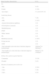
Baseline characteristics of the patients.
| Patient baseline characteristics | N (%) |
| Sex | |
| Male | 8 (42) |
| Female | 11 (58) |
| Age (range) | 2.1 years (0.25–14) |
| Underlying disease | |
| ALL | 7 (36) |
| AML | 2 (10.5) |
| Atypical choroid plexus papilloma | 1 (5) |
| Choroid plexus carcinoma | 1 (5) |
| Medulloblastoma | 1 (5) |
| PNET | 1 (5) |
| Wilms’ tumour | 1 (5) |
| HL | 1 (5) |
| Rhabdomyosarcoma | 2 (10.5) |
| Ewing sarcoma | 1 (5) |
| SCID | 1 (5) |
| Total neutrophil count at the time of infection diagnosis (median/range) | 1500/mm3 (0–9600/mm3) |
| Total lymphocyte count at the time of infection diagnosis (median/range) | 940/mm3 (110–3700/mm3) |
| Setting where patient acquired infection | |
| Community | 15 (79) |
| Hospital | 4 (21) |
| Duration of symptoms prior to first visit | |
| 1 day | 12 (63) |
| 2 days | 3 (16) |
| 3 days | 4 (21) |
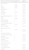
Presenting characteristics of the RSV infection and its complications.
| Characteristics of RSV infection in patients | Not admitted (10; 53.7%) | Admitted (9; 47.3%) |
| Signs and symptoms | ||
| Fever | 8 (80%) | 9 (100%) |
| Cough | 10 (100%) | 7 (77.7%) |
| Rhinorrhoea | 7 (70%) | 7 (77.7%) |
| Respiratory distress | 0 | 2 (22.2%) |
| Wheezing | 0 | 2 (22.2%) |
| Arthromyalgia | 1 (10%) | 1 (11.1%) |
| Hypoxaemia | 0 | 2(22.2%) |
| Chest radiography at diagnosis (10 patients) | 4 (40%) | 6 (66.6%) |
| Normal | 4 (100%) | 2 (22.2%) |
| Bilateral hilar infiltrates | 0 | 1 (11.1%) |
| Bilateral consolidations | 0 | 1 (11.1%) |
| Acute respiratory distress syndrome | 0 | 1 (11.1%) |
| Mediastinal mass | 1 (10%) | 0 |
| Report was unavailable | 0 | 1 (11.1%) |
| Requiring admission to PICU | ||
| Yes | 1 (11.1%) | 2 (22.2%) |
| No | – | 7 (77.7%) |
| Complications of RSV infection | ||
| Acute respiratory distress syndrome | 0 | 2 (22.2%) |
| Heart failure | 0 | 1 (11.1%) |
| Pulmonary haemorrhage | 0 | 1 (11.1%) |
| Pneumothorax | 0 | 1 (11.1%) |
| Hypertransaminasaemia | 0 | 1 (11.1%) |
| Hospital-related complications | ||
| Port-a-cath infection | – | 3 (33.3%) |
| Urinary tract infection | – | 1 (11.1%) |
| Severe withdrawal symptoms | – | 2 (22.2%) |
| Malnutrition | – | 2 (22.2%) |
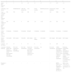
Summary of the impact in length of stay in days, respiratory support, and specific treatment in each patient.
| 1 | 2 | 3 | 4 | 5 | 6 | 7 | 8 | 9 | |
| Age at RSV diagnosis (years | 4 | 2 | 1.9 | 1.6 | 12 | 1.5 | 1.5 | 0.25 | 1.6 |
| Underlying disease | ALL | Medulloblastoma | AML/2nd tumour ALL | ALL | Ewing sarcoma | Rhabdomyosarcoma | Promyelocytic leukaemia | SCID | ALL |
| Total neutrophil count at time of RSV diagnosis (/mm3) | 500 | 700 | 3100 | 290 | 30 | 2225 | 160 | 5600 | 1500 |
| Total lymphocyte count at time of RSV diagnosis (/mm3) | 700 | 700 | 0 | 940 | 110 | 1.700 | 140 | 700 | 400 |
| Setting where infection was acquired | Community | Community | Community | Hospital | Community | Community | Community | Community | Community |
| Method used to diagnose infection | Viral culture | RSV antigen | Viral PCR | RSV antigen | RSV antigen | Viral culture | RSV antigen | RSV antigen | RSV antigen |
| Length of stay (days) | 20 | 35 | 4 | 34 | 6 | 4 | 10 | 100 | 50 |
| Associated pathology | Infectious papular skin lesions of undetermined source. Pancytopaenia following chemotherapy. | Fever and neutropaenia following chemotherapy | Fever and neutropaenia | Escherichia coli urinary tract infection. Port-a-cath infection by Staphylococcus epidermidis | Fever and neutropaenia | No | Fever and neutropaenia | ARDS, pneumothorax, heart failure, coinfection by Pneumocystis jiroveci, severe withdrawal syndrome. Severe malnutrition. | Pulmonary haemorrhage, ARDS, pneumothorax, haemothorax, haemorrhagic shock, coagulate-negative Staphylococcus bacteraemia, Corynebacterium bacteraemia |
| Respiratory support | – | – | – | – | – | – | – | HFOV, PMAP 24cm H2O, peak amplitude 55cm H2O, frequency 8Hz, FiO2 0.9 | HFOV, FiO2 100%, Paw 31, A 75, Ti (%) 33; and frequency 10Hz |
Chemotherapy had to be delayed due to RSV infection in 4 patients (for 7 days in 3 patients, and 29 days in 1 patient).
There were no fatalities associated to RSV infection in the cohort during the period of the study.
DiscussionInfection by RSV can become serious in immunocompromised patients,6 as demonstrated by the 2 patients in our cohort that required admission to the PICU and respiratory support (the patients admitted to the PICU were those who needed respiratory support beyond basic oxygen therapy), with a proportion similar to those observed in other studies.7 A greater severity in these infections has been correlated with a greater degree of immunosuppresion,4 as was the case of the 2 patients admitted to the PICU. Furthermore, immunocompromised patients need to be admitted to the hospital more frequently, which may in turn lead to nosocomial complications (Tables 2 and 3). Treatments of RSV of demonstrated efficacy in immunocompromised patients include antiviral agents such as ribavirin; palivizumab; and immune globulin therapy.8–10 In our cohort, oral ribavirin was administered to one patient with SCID that developed ARDS secondary to RSV infection. Ribavirin is a nucleoside analogue with antiviral activity against RNA viruses. Administration of inhaled ribavirin requires specific environmental precautions (administration in a room with negative air pressure and use of protective masks) and has been associated with respiratory side effects.11 Oral administration has been proven to be safe and equally efficacious.12 However, it has been hypothesised that oral administration may not prevent progression of the infection, and consequently suggested that treatment starts with a high dose that is later escalated according to how the patient responds.13
The appropriate duration of treatment has yet to be established, although most groups continue pharmacological therapy until the virus is cleared. The combination of antiviral agents with the monoclonal antibody palivizumab is safe, well-tolerated by the patient, and has shown high efficacy in association with symptom resolution and viral clearance.14
Supportive care is essential to ensure that patients receive the necessary nutrition, hydration, and respiratory support, and ECMO is particularly important in the management of refractory hypoxaemia in patients that develop ARDS secondary to infection with RSV and do not respond adequately to conventional mechanical ventilation or HFOV.15 In patients receiving an HSCT, delaying chemotherapy has been associated with a lower rate of progression to pneumonia.16 Future research could investigate how delaying treatment of the underlying disease as a result of RSV infection impacts the overall survival rate of patients. In our cohort, 21% of RSV infections were nosocomial. When a case of RSV is detected, isolation measures must be taken, handwashing practised conscientiously, and the rest of the patients must be watched carefully for infection with RSV.16 Some research groups propose that palivizumab be administered to hospitalised patients as a prophylactic measure to prevent an outbreak among immunocompromised patients when a case of RSV is first detected.17
Infection by RSV in immunocompromised patients is associated with high morbidity and mortality, as seen in the 2 patients in our series that required admission to the PICU. Survival in our series reached 100%, which highlights the importance of maintaining a high degree of vigilance for these infections, the early initiation of supportive care and specific treatment, and implementing isolation measures to control infection and prevent outbreaks.
Supportive care measures are key in the management of these patients, and ECMO in particular in patients that develop severe complications such as ARDS or pulmonary haemorrhage.
Conflicts of interestThe authors have no conflicts of interest to declare.
Please cite this article as: Domínguez-Pinilla N, Belda Hofheinz S, Vivanco Martinez JL, Baro-Fernández M, Ruiz-Contreras J, González-Granado LI. Infección por virus respiratorio sincitial en los pacientes inmunodeprimidos en un hospital pediátrico: experiencia de 5 años. An Pediatr (Barc). 2015;82:35–40.
