Información de la revista
Vol. 100. Núm. 2.
Páginas 153-154 (Febrero 2024)
Images in Paediatrics
Acceso a texto completo
A rare cause of bronchiectasis
Bronquiectasias de causa infrecuente
Visitas
2509
Carlos Martín de Vicente
Autor para correspondencia
clmartin@salud.aragon.es

Corresponding author.
, Mercedes Odriozola Grijalba
Unidad de Neumología Pediátrica, Hospital Universitario Miguel Servet, Zaragoza, Spain
Este artículo ha recibido
Información del artículo
Texto completo
Bibliografía
Descargar PDF
Estadísticas
Figuras (3)
Mostrar másMostrar menos
Material adicional (1)
Texto completo

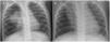
A girl aged 6 years with oculocutaneous albinism type 3 had pneumonia in the right upper lobe (RUL) of the lung at birth, and developed persistent infiltrate in the same location at a later time (Fig. 1). A computed tomography (CT) scan of the lung evinced partial atelectasis of the RUL with cystic bronchiectasis in the apical and anterior segments, right aortic arch and dilatation of the pulmonary artery (Fig. 2). Flexible bronchoscopy allowed the detection, near the carina, of a tracheal bronchus into which the bronchoscope could not advance due to collapse of the lumen, protrusion of the mucosa at the start of the right main bronchus and a decreased caliber of the opening to the RUL, with detection of two obliterated lumina (anterior and posterior segments) (Appendix A, Video 1).

Figure 1.

Persistent infiltrate in the right upper lobe: (A) age 1 year; (B) age 2 years.

Figure 2.

Bronchiectasis in the right upper lobe: (A) axial plane; (B) coronal plane; (C) sagittal plane; (D) right aortic arch.

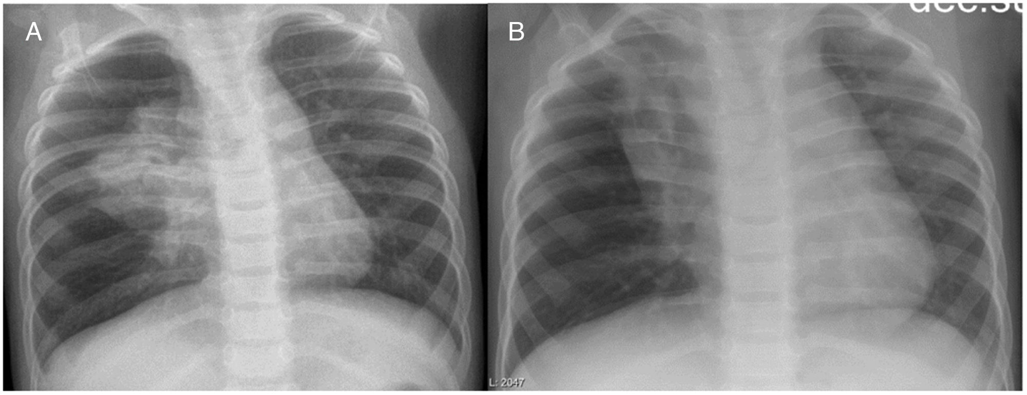
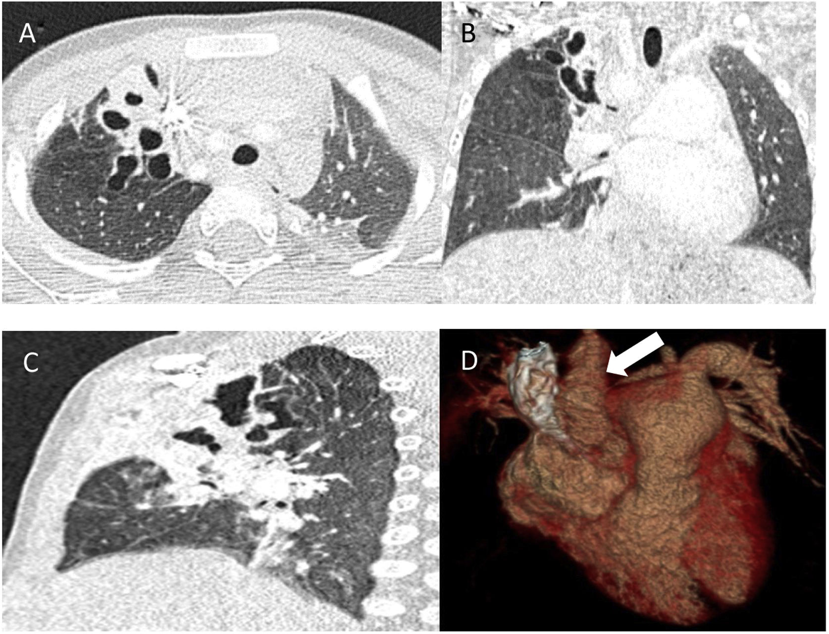
These findings suggested a tracheobronchial malformation due to the displacement of the apical segment of the RUL bronchus1 and the compression of this segment and the anterior segment by the aorta, with an abnormal right location, and development of bronchiectasis, which is not usually associated with tracheal bronchus.2,3 Due to the favourable clinical course, a conservative watchful waiting approach was decided on, and the patient is currently free of pulmonary infections. The follow-up CT scan 4 years later revealed residual retractile atelectasis with a fibrous appearance and bronchiectasis (Fig. 3).

Figure 3.

Follow-up computed tomography (CT) scan of the lungs (4 years later).

Funding

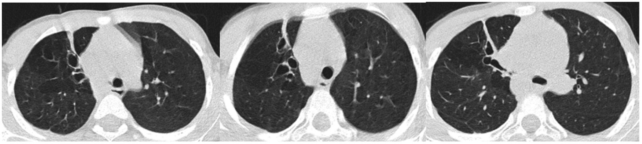
This research did not receive any external funding.

Appendix A
Supplementary data

The following is Supplementary data to this article:

(2.71MB)

References
[1]
J.L. Pérez, S. Caussade.
Bronquio traqueal.
Neumol Pediatr, 7 (2012), pp. 58-60
[2]
A. Jamil, A. Jamil, A. Popa, H. Ismail.
Tracheal bronchus: rare cause of recurrent pneumonia and worsening bronchiectasis in adults.
Clin Med Rev Case Rep, 6 (2019), pp. 277
[3]
M. Moreno, S. Castillo-Corullón, E. Pérez-Ruiz, M.C. Luna, J.L. Antón-Pacheco, P. Mondejar-Lopez, et al.
Spanish multicentre study on morbidity and pathogenicity of tracheal bronchus in children.
Pediatr Pulmonol, 54 (2019), pp. 1610-1616
Copyright © 2023. Asociación Española de Pediatría
Descargar PDF
Idiomas
Anales de Pediatría
Opciones de artículo
Herramientas
Material suplementario