Sugerencias
Compartir
Información de la revista
Vol. 98. Núm. 6.
Páginas 481-482 (Junio 2023)
Images in Paediatrics
Acceso a texto completo
Congenital lobar emphysema simulating a pneumothorax
Enfisema lobar congénito simulando un neumotórax
Visitas
4050
Luis Vega Lópeza,
Autor para correspondencia
Luisvegalopez11@hotmail.com

Corresponding author.
, Sonia Lareu Vidalb, Gonzalo Anes Gonzálezc, Víctor Álvarez Muñozd
a Servicio de Pediatría, Hospital Universitario Central de Asturias, Oviedo, Spain
b Sección de Neonatología, Servicio de Pediatría, Hospital Universitario Central de Asturias, Oviedo, Spain
c Servicio de Radiodiagnóstico, Hospital Universitario Central de Asturias, Oviedo, Spain
d Servicio de Cirugía Pediátrica, Hospital Universitario Central de Asturias, Oviedo, Spain
Este artículo ha recibido
Información del artículo
Texto completo
Bibliografía
Descargar PDF
Estadísticas
Figuras (3)
Mostrar másMostrar menos
Material adicional (1)
Texto completo

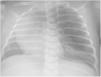
A neonate was referred to our hospital at 13 days post birth for assessment of dyspnoea. In the referring hospital, the complete blood count results had been normal, while the blood gas analysis revealed respiratory acidosis and the chest radiograph findings (Fig. 1) were compatible with pneumothorax.

Figure 1.

Hyperinflated and hyperlucent left hemithorax with contralateral mediastinal shift.

However, the physical examination did not evince the expected severity, finding a mild increase in the work of breathing and cyanotic spells limited to intense crying.

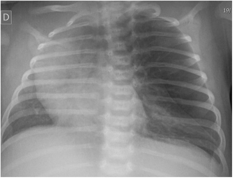
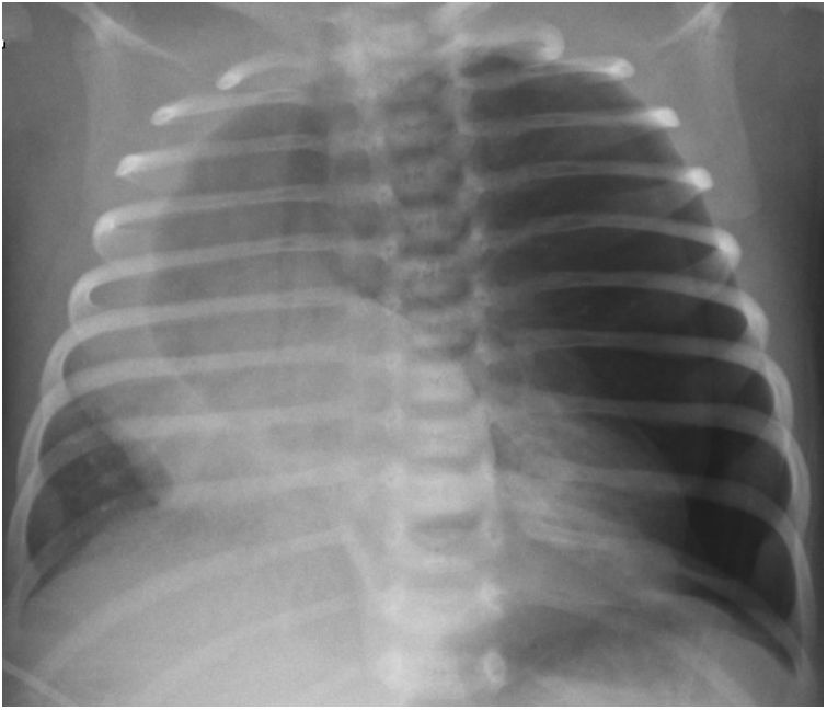
After drainage, the condition of the infant worsened abruptly with shifted cardiac sounds and marked hypoventilation of the affected hemithorax. A new chest radiograph (Fig. 2) detected a tension pneumothorax in addition to the pre-existing lesion.

Figure 2.

Iatrogenic pneumothorax in left lung associated with the pre-existing congenital lobar emphysema with marked contralateral mediastinal shift.

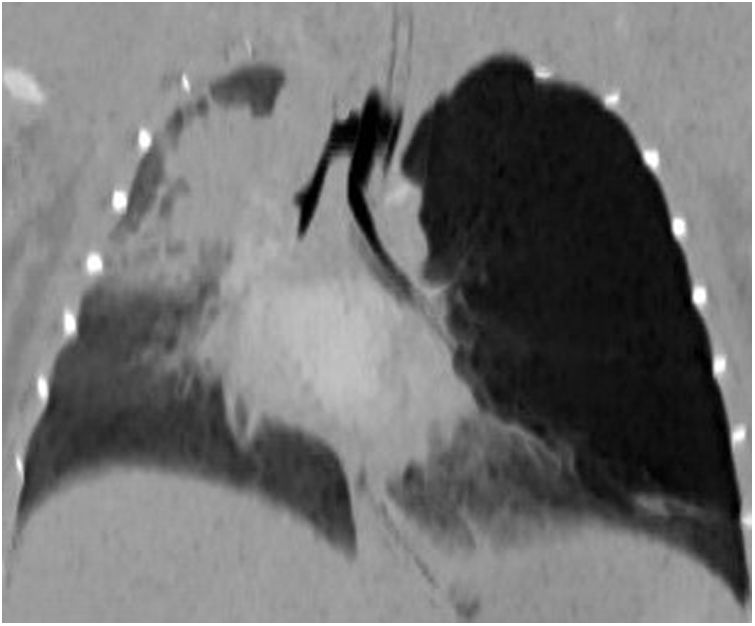
The change in the patient’s condition prompted the performance of a computed tomography scan (Fig. 3; Appendix B, video 1), which resulted in the definitive diagnosis of congenital lobar emphysema.

Figure 3.

Coronal minimum-intensity projection. Hyperlucent area with mass effect in the left upper lobe and contralateral mediastinal shift. Right upper lobe atelectasis.

At the time of this writing, after undergoing lobectomy via thoracotomy in the department of paediatric surgery, the patient is asymptomatic, and the check-ups performed to date have evinced normal development.

Congenital lobar emphysema is an infrequent disease caused by alterations in lung development that result in air trapping through a “ball-valve” mechanism.1

At the time of diagnosis, it is common to mistake congenital lobar emphysema with pneumothorax,2,3 so it is important to take the former into account in the differential diagnosis and perform additional tests as needed. Although the sonographic features of this disease have yet to be established, a lung ultrasound scan could have been useful in this case, as it would not have shown the characteristic pattern of pneumothorax.

Appendix A
Supplementary data

The following is Supplementary data to this article:

(11.02MB)

References
[1]
Oermann CM. Congenital lobar emphysema. UptoDate. 2021 [accessed 16 May 2022]. Available from: https://www.uptodate.com/contents/congenital-lobar-emphysema?search=congenital%20enfisema&source=search_result&selectedTitle=1∼150&usage_type=default&display_rank=1.
[2]
S.M. Prabhu, S.R. Choudhury, R.S. Solanki, G.S. Shetty, S. Agarwala.
Inadvertent chest tube insertion in congenital cystic adenomatoid malformation and congenital lobar emphysema-highlighting an important problem.
[3]
A. Reed, S.F. Lucas, A. Nowacka, C. Eze.
Iatrogenic pneumothorax in a 4-week-old girl: new diagnosis of congenital lobar emphysema.

Previous meeting: this case was presented at the Spring Meeting of the Sociedad de Pediatría de Asturias, Cantabria y Castilla y León (SCCALP); April 29-30, 2022; Avilés, Spain.

Copyright © 2022. Asociación Española de Pediatría
Descargar PDF
Idiomas
Anales de Pediatría
Opciones de artículo
Herramientas
Material suplementario