


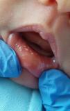
A full-term male neonate born via elective caesarean section had an uneventful antenatal and perinatal course, with the exception of maternal colonization by group B Streptococcus, which was managed with intrapartum prophylaxis. On the third day of life, multiple millimetric whitish papulovesicular lesions were observed inside the lower lip (Fig. 1). The remainder of the physical examination was unremarkable. The patient received a clinical diagnosis of intraoral sebaceous hyperplasia. Spontaneous regression was reported three days later.

Sebaceous gland hyperplasia is a common neonatal finding. However, the intraoral presentation, referred to as Fordyce granules, is rare, with an estimated incidence of 1%.1 It is a benign condition characterized by the presence of ectopic sebaceous glands, believed to result from maternal androgenic stimulation during the final month of gestation and associated with the maturity of the newborn.1,2 The diagnosis is clinical, based on the identification of small, asymptomatic, whitish or yellowish papulovesicular lesions that may coalesce.2 Differentiation from pathological conditions is essential, particularly from infection by herpes simplex virus, which is rarely limited to the oral mucosa and is usually associated with systemic involvement and ulcerative lesions.2,3 This disease is self-limiting and usually resolves spontaneously.2 Early recognition is important to prevent unnecessary procedures and to reassure parents.1
