


Strength is a physical quality with a clear influence on quality of life. It is determined by the structure of the musculoskeletal system, and depends on the muscular structure. It has been described that prematurity conditions both qualities. The aims of this study are to determine whether prematurity is associated with strength or body composition and to evaluate the relationship between prematurity, strength and muscle mass.
Material and methodsCase–control study. Participants were premature 7-to-11-year-old children and full-term birth controls. Strength was measured by a strength gauge and body composition from DEXA (duel-energy X-ray absorptiometry) scans. A total of 89 subjects were included and divided into three groups: 30 prematures with birth weight ≤1500g, 29 prematures with birth weight 1500–2000g, and 30 controls.
ResultsWeight and BMI z-score was lower in the premature group. No differences were found in muscular mass or strength between groups. A ratio was established between strength and weight or muscular mass. It was observed that it was possible for them to move four times their weight, without finding any differences between groups or a relationship with birth weight.
ConclusionsBetween 7 and 11 years of age, children who were premature have lower weight and BMI than the rest of the children. However, there were no differences in body composition or strength between preterm children and controls.
La fuerza es una cualidad con clara influencia sobre la calidad de vida. Está condicionada por la estructura del aparato locomotor y es directamente dependiente de la estructura muscular. Se ha descrito que ambas cualidades están condicionadas por la prematuridad. Son objetivos del estudio conocer si la prematuridad está relacionada con la fuerza o la composición corporal durante la infancia y valorar la relación entre prematuridad, fuerza y masa muscular.
Material y métodosEstudio de casos y controles realizado en niños de entre 7 y 11 años con desarrollo normal y controles a término de la misma edad. Se incluyó a 89 sujetos: 30 prematuros con peso al nacimiento ≤ 1.500g, 29 prematuros con peso al nacimiento > 1.500g y 30 controles. Se analizaron antropometría, composición corporal mediante absorciometría de rayos X de energía dual y fuerza isométrica mediante banco inclinado y galga.
ResultadosEl peso y el IMC fueron menores en los niños que pesaron ≤ 1.500g. No se observaron diferencias en composición corporal ni fuerza. Se estableció una razón entre fuerza y masa muscular, resultando esta de un peso desplazado 4 veces superior al peso corporal, no encontrándose diferencias entre grupos ni relación con el peso al nacimiento.
ConclusionesEntre los 7 y los 11 años de edad, los niños que fueron grandes prematuros tienen un peso y un IMC menores al resto de los niños. No se encontraron diferencias entre prematuros y controles en cuanto a composición corporal y fuerza muscular.
Neonatal care has become specialised and improved considerably, progressively incorporating new technologies and scientific advances.1 This progress has been most evident in the premature population. The number of preterm and very preterm patients has increased considerably in recent decades.2
The body composition of individuals changes over time during childhood and adolescence.3,4 In addition to a quantitative evolution that is parallel to growth, continuous changes in the proportions and characteristics of its components take place.5 Furthermore, in the past two decades, skeletal muscle has been identified as a fundamental component of the immune system and as an endocrine organ.6
Strength is defined as the basic physical ability that allows us to generate muscle tension in single maximal effort to overcome a resistance or load.7 It is conditioned by the structure of the musculoskeletal system and determined in part by muscular structure.
Some authors have stated that the level of physical activity of individuals born preterm is lower than that of individuals born at term.8,9 Similarly, it has been reported that overall, individuals born preterm have more muscle tone and motor coordination problems,10–12 and that their muscle strength is lesser in many instances.13
Confirmation that these deficits exist in premature children and the degree to which they impact them would allow for the early implementation of strategies aimed at minimising these deficits, optimising the available resources and improving the prognosis of this population.
The aim of this study was to determine whether prematurity is associated with body composition and strength during childhood, and to assess the association between prematurity, strength, and lean mass or muscle mass.
Materials and methodsWe conducted an observational, cross-sectional, descriptive and prospective case–control study.
SampleWe included children born between January 1, 2001 and December 31, 2004. The data were collected throughout 2012. Thus, study participants were 7–11 years of age.
A total of 89 participants, 37 male and 52 female, were included in the study.
CasesChildren born preterm who were admitted to the neonatal unit and had a seemingly normal psychomotor and cognitive development were studied. We defined prematurity as birth occurring before the start of the 37th week of gestation. We subsequently divided the cases into two groups based on birth weight, PREM_1 (birth weight≤1500g) and PREM_2 (birth weight>1500g). The sex distribution was random. Corrected age was calculated based on 40 weeks of gestation.
Matched controlsThe controls were healthy individuals born at term. The number and age of the controls had to be similar to those of the cases. We refer to these participants as the control group.
Exclusion criteriaWe excluded any children diagnosed with diseases that manifest with psychomotor or cognitive deficits.
MethodsMedical chart reviewWe collected data on anthropometric measurements at birth and any complications during the stay in the neonatal unit.
Anthropometric measurementsParticipants were weighed and their height was measured by dual-energy X-ray absorptiometry (DXA). We calculated the body mass index (BMI) for each participant. To correct for potential age- or sex-related biases, we calculated the z-score for weight, height and BMI with the following formula: z-score=(measurement−mean)/standard deviation.
We located the mean±standard deviation for each measurement in the growth charts developed by Carrascosa et al.14
Determination of body compositionDual X-ray absorptiometry is a reliable method for assessing body composition, estimating the bone mineral density, fat mass and muscle mass.
Body composition was assessed with a DXA scan using the General Electric Lunar Prodigy Pro® analyser and the EnCORE 2009® operator platform. Lean mass, fat mass and bone mass were assessed by whole-body scans with the patient placed supine in the anatomical position. The software of the device allowed us to select the lean mass of the lower limbs.
Strain gaugeThe strain gauge, adapted to different devices so that force can be exerted on it, can be used to measure isometric force.
The maximal static strength of the lower body of the participants was measured using a 45° Gerva-Sport® incline leg press (Spain) (Fig. 1) fitted with a Globus Ergometer® strain gauge or load cell that was in turn connected to an Ergo Tester® v 1.5 microprocessor (Italy) (Fig. 2), downloading the data with the TAL Technologies Software Graph® application.


Participants were asked to sit on the press, placing their feet on the platform in parallel and shoulder-width apart. Their knees had to be flexed forming a 110° angle between leg and thigh (Fig. 3). Once the child was positioned, there was a three-second countdown after which the participant had to push the platform as hard as possible from the start and try to hold its position for a full five seconds. Following a brief explanation of the apparatus and technique by the researcher, the measurement with the gauge strain fitted to the incline leg press could be performed correctly in nearly every participant.
Statistical analysis
We used Microsoft Excel 2010 and the statistical software SPSS V18.0. We compared qualitative variables by means of the chi-square test, and quantitative variables with parametric tests (Student t test) when the sample followed a normal distribution or nonparametric tests when the distribution was not normal. We analysed significance by multiple factor ANOVA, assessing for linear and nonlinear correlations.
Ethical aspectsThe privacy of participants was protected in accordance with the 2008 Declaration of Helsinki on informed consent and research involving animals and human beings.15 Participation in the study required the signing of an informed consent form designed specifically for the purpose.
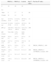
ResultsSample characteristicsTable 1 summarises the characteristics of the participants, including age, corrected age, birth weight and sex distribution.
Age, sex and anthropometric characteristics of the groups under study.
| PREM_1 | PREM_2 | Control | Sig (P value) | Post hoc(P value) | |
|---|---|---|---|---|---|
| N | 30 | 29 | 30 | ||
| Sex | |||||
| Male | 9 | 17 | 11 | .06 | |
| Female | 21 | 12 | 19 | ||
| Age (months) | 113.53±15.45 | 111.10±12.42 | 107.23±15.65 | .14 | |
| Corrected age | 111.40±15.50 | 110.38±12.13 | 107.23±15.65 | .56 | |
| BW (g) | 1261.40±255.15 | 1822.14±199.57 | 3231.23±398.81 | .000 | |
| BW (range) | 720–1500 | 1570–2374 | 2495–3860 | ||
| Weight (kg) | 30.0±8.2 | 34.5±9.4 | 32.5±8.8 | .15 | |
| Height (cm) | 132.7±9.7 | 133.9±9.1 | 134.3±10.7 | .81 | |
| BMI (kg/m2) | 16.7±2.6 | 18.9±3.3 | 17.7±2.7 | .02 | PREM_1/PREM_2: .006 |
| Weight z-score | −0.6±1.2 | 0.3±1.3 | 0.3±1.1 | .008 | PREM_1/PREM_2: .007PREM_1/control: .009 |
| Height z-score | −1.1±4.4 | 0.1±1.5 | 0.6±1.7 | .083 | |
| BMI z-score | −0.6±1.4 | 0.3±1.1 | 0.0±0.8 | .007 | PREM_1/PREM_2: .002PREM_1/control: .036 |
BMI, body mass index; BW, birth weight; control, control group; PREM_1, group of preterm individuals with BW ≤1500g; PREM_2, group of preterm individuals with BW 1500–2500g; Sig, statistical significance.
We observed that the groups were comparable in terms of age and corrected age. The number of participants in each group was also similar.
Anthropometric characteristics are also summarised in Table 1. We observed that weight and height were similar in both groups, and we found no statistically significant differences between them for these variables. However, we found that the BMI was lower in the PREM_1 group. This difference was more marked when we analysed the weight and BMI z-scores, which were lower in the PREM_1 group; the difference reached statistical significance.
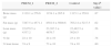
Body compositionTable 2 shows the amount of bone mass, lean mass and fat mass in each group, estimated by DXA. They were similar in both groups and the differences we found were not statistically significant.
Body composition in lean tissue mass, fat tissue mass and bone mass of the groups under study.
| PREM_1 | PREM_2 | Control | Sig (P value) | |
|---|---|---|---|---|
| Bone mass (g) | 1119.1±276.6 | 1154.1±265.8 | 1115.1±359.8 | .87 |
| Fat mass (g) | 7287.5±4571.1 | 8581.9±5600.6 | 7632.4±5217.5 | .62 |
| Lean mass (g) | 22,079.5±4337.2 | 23,387.3±4676.7 | 22,482.5±5020.5 | .56 |
| % fat | 22±10 | 24±10 | 23±10 | .78 |
| % lean tissue | 74±9 | 73±10 | 74±10 | .80 |
Control, control group; PREM_1, group of preterm individuals with BW ≤1500g; PREM_2, group of preterm individuals with BW 1500–2500g; Sig, statistical significance.
We also present the percentage of the total body mass constituted by lean mass and fat mass in each group. We found no statistically significant differences between both groups.
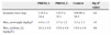
Isometric strength in the lower limbsTable 3 shows the isometric strength of the lower limbs that was measured in the leg press expressed as kilograms.
Expressions of strength assessed with the strain gauge.
| PREM_1 | PREM_2 | Control | Sig (P value) | |
|---|---|---|---|---|
| Isometric force (kg) | 115.2±34.4 | 115.5±35.1 | 109.90±38.5 | .82 |
| Max_iso/weight (kg/kg)a | 4.0±1.5 | 3.7±1.3 | 3.5±1.1 | .43 |
| Max_iso/lean_LL (kg/kg)b+ | 16.2±4.8 | 15.6±4.6 | 15.0±4.8 | .68 |
Control, control group; max_iso, isometric force in lower limbs; lean_LL, lean mass in the lower limbs; PREM_1, group of individuals born preterm with birth weight ≤1500g; PREM_2: group of individuals born preterm with birth weight 1500–2500g; sig, statistical significance.
The isometric force exerted in the incline press was similar in all groups, and we found no statistically significant differences.
The mean ratios of the isometric force on the incline press and the total body mass and of the isometric force and the lean mass of the lower limbs, which were expressed as kilograms of force per kilograms of body mass or lean mass of the lower limbs (kg/kg), are presented in Table 3. The isometric force exerted relative to the body mass or the lean mass of the lower limbs was similar in all groups. Generally, participants exerted a force that was four times their body mass. We did not find statistically significant differences between groups.
We did not find a correlation between birth weight and the mean ratio of the isometric force exerted on the press and the lean mass of the lower limbs.
DiscussionIndividuals engage in physical activity throughout their lives. Movement starts even before birth, during gestation. Previously acquired abilities improve significantly between 6 and 14 years of age, before adolescence.
Many characteristics influence physical activity and consequently quality of life. Some physical qualities have a decisive influence, including strength, endurance, flexibility or speed. Body composition is another determinant closely related to those just mentioned.
The sample selected based on the inclusion criteria established for the study turned out to be adequate. The case and control groups were similar both in the number and ages of their members and in their age composition. The sex distribution was random and different in the case and control groups. However, we did not take this into account, as sexual dimorphism in strength and body composition gains significance mostly from 12 years of age onward.16
Body compositionMultiple factors influence body composition in adulthood. The catch-up growth17,18 experienced by individuals born preterm may promote a greater percentage of fat mass. However, fat mass is associated more strongly with adult weight than with birth weight. The same is true of lean mass, which is proportionally lesser in subjects that are heavier in adulthood despite being greater in absolute terms.
In our study, the analysis of anthropometric measurements revealed a lower body weight and BMI in individuals born very preterm. This finding is consistent with the literature.17,19,20
There is published evidence that birth weight does not influence body composition in adulthood.9,21,22 Our study attempted to establish whether it influenced body composition in school-aged children. Furthermore, determining the body composition is important in the assessment of strength. We assessed body composition by means of DXA. Its use in the paediatric age group has increased in recent years. The radiation the patient is exposed to is less than a tenth of the radiation of a chest radiograph, and is much lower than the radiation produced by other tests that are used frequently.23–30
DXA was performed adequately on every study participant. We found no differences between the premature group and the control group in fat mass, bone mass or lean mass. This finding is consistent with the literature, with studies analysing the differences in adolescents19 and most frequently young adults.9,18,21,22 That is, in the participants in our sample, aged 7–11 years, body composition was similar irrespective of birth weight and the differences found in weight and BMI.
Muscle strength and qualityStrength is a physical quality that has a clear influence on the quality of life of individuals. There are several types of strength. Maximum strength is that which overcomes a maximal resistance. Explosive strength or power is the strength involved in overcoming a small resistance with maximal speed. Endurance strength is the ability to perform the muscular effort repeatedly for a long time.
In this study, we assessed the maximal isometric force of the lower limbs. In absolute terms, the exerted force was similar across groups.
There is published evidence supporting a correlation between muscle function in individuals born preterm and the degree of prematurity, anthropometric measurements at birth, and auxological characteristics. Some studies have also observed that individuals born preterm have less strength than individuals of the same age born at term.13,31,32 This difference is more marked in preterm individuals that experienced complications during the neonatal period. Unlike the study we are presenting, most past studies have been conducted in the adult or adolescent population,8 and most used other methods for strength assessment. The difference in the dates of birth, which ranges approximately from one to three decades due to both the age of the participants and the year the studies were conducted, may have determined differences in care during the neonatal period given how much the field of neonatology has advanced both at the technical and the scientific levels. Studies conducted on children31 have had narrower participant age ranges and used other methods for lower limb strength assessment.
We assessed the relationship between the exerted force and participant characteristics. When we assessed it in relation to the body mass of participants by dividing the exerted force by the body weight, we observed that participants exerted a force equivalent to about four times their weight. This result was similar in every group, with the proportion remaining the same. We also studied the ratio between isometric force and the lean mass of the lower limbs, as we believed that this would be an adequate variable for muscle function assessment. We observed that the ratio was maintained across all groups, which demonstrates that muscle strength is similar in individuals born preterm and individuals born at term.
Thus, with the instruments used in this study we found that, unlike what had been observed in previous studies, the strength measured in individuals born preterm and individuals born at term was similar in our sample. As mentioned above, participants in this study were born more recently, which may have influenced the care they received at the hospital. Furthermore, most of the previous studies were conducted in young adults.8,13,32 While we assume that the characteristics of the participants in our sample will hold in the future, follow-up of the sample for confirmation should be considered. Lastly, this is the first time that a strain gauge attached to an incline leg press was used to assess isometric strength in the lower limbs of individuals born preterm. Previous studies had used methods based on simple exercises, and most importantly, had assessed strength in the upper limbs.
ConclusionsBased on the results of this study, differences in muscle strength or body composition between children born preterm and children born at term cannot be established using DXA scans for the assessment of body composition and a strain gauge fitted to an incline press for measuring maximal isometric force in the lower limbs.
We also did not find differences between individuals born preterm and individuals born at term when we calculated the force to lean mass ratio to assess muscle function.
We observed a lower weight and BMI in the very preterm group. These findings are consistent with the literature, as catch-up growth is not complete in the age range under study.
FundingThis study is part of a larger project that has received one of the XIV grants for clinical and epidemiological research of the Fundación Ernesto Sánchez Villares (2011).
Conflicts of interestThe authors have no conflicts of interest to declare.
Please cite this article as: Mata Zubillaga D, Rodríguez Fernández C, Rodríguez Fernández LM, de Paz Fernández JA, Arboleda Franco S, Alonso Patiño F. Valoración de fuerza isométrica en extremidades inferiores y composición corporal en prematuros. An Pediatr (Barc). 2015;83:229–235.
