
Multiple sclerosis is the leading cause of severe neurologic impairment in young individuals, resulting in significant disability. Although the disease is infrequent in the paediatric population, it is estimated that the onset of symptoms occurs before age 18 years in 10% of cases.
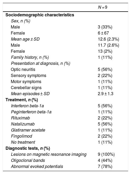
We present a series of 9 cases of multiple sclerosis diagnosed based on the McDonald criteria in the Neuro-Ophthalmology Unit of the Hospital Clínico San Carlos of Madrid, Spain. We found a higher proportion of female patients, who were also diagnosed at an older age (Table 1). The predominant presentation was optic neuritis and sensory symptoms in female patients and motor symptoms in male patients. Forty-four percent of patients had a second episode within a year. The visual acuity of patients that had onset with optic neuritis was greater than 20/30. Optical coherence tomography did not reveal thinning of the retinal nerve fibre layer, but there was evidence of a decreased P100 amplitude with a prolonged latency in the visual evoked potentials.
Characteristics of patients with juvenile multiple sclerosis.
| N = 9 | |
|---|---|
| Sociodemographic characteristics | |
| Sex, n (%) | |
| Male | 3 (33%) |
| Female | 6 ± 67 |
| Mean age ± SD | 12.6 (2.3%) |
| Male | 11.7 (2.6%) |
| Female | 13 (2%) |
| Family history, n (%) | 1 (11%) |
| Presentation at diagnosis, n (%) | |
| Optic neuritis | 5 (56%) |
| Sensory symptoms | 2 (22%) |
| Motor symptoms | 1 (11%) |
| Cerebellar signs | 1 (11%) |
| Mean episodes ± SD | 2.9 ± 1.3 |
| Treatment, n (%) | |
| Interferon beta-1a | 5 (56%) |
| Peginterferon beta-1a | 1 (11%) |
| Rituximab | 2 (22%) |
| Natalizumab | 5 (56%) |
| Glatiramer acetate | 1 (11%) |
| Fingolimod | 2 (22%) |
| No treatment | 1 (11%) |
| Diagnostic tests, n (%) | |
| Lesions on magnetic resonance imaging | 9 (100%) |
| Oligoclonal bands | 4 (44%) |
| Abnormal evoked potentials | 7 (78%) |
SD, standard deviation.
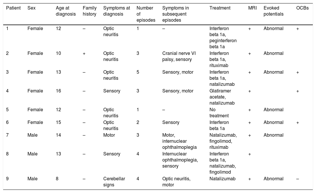
To date, all the patients have had a relapsing-remitting course. Four patients received natalizumab as first-line treatment because they had 2 or more episodes in the first year. Two patients received rituximab as second-line treatment after failure of interferon beta and fingolimod. Both patients tested positive for Creutzfeldt-Jakob disease, so treatment with natalizumab was not contemplated (Table 2).
Clinical characteristics, outcomes and diagnostic tests in patients given a diagnosis of multiple sclerosis.
| Patient | Sex | Age at diagnosis | Family history | Symptoms at diagnosis | Number of episodes | Symptoms in subsequent episodes | Treatment | MRI | Evoked potentials | OCBs |
|---|---|---|---|---|---|---|---|---|---|---|
| 1 | Female | 12 | – | Optic neuritis | 1 | – | Interferon beta 1a, peginterferon beta 1a | + | Abnormal | + |
| 2 | Female | 10 | + | Optic neuritis | 3 | Cranial nerve VI palsy, sensory | Interferon beta 1a, rituximab | + | Abnormal | |
| 3 | Female | 13 | – | Optic neuritis | 5 | Sensory, motor | Interferon beta 1a, natalizumab | + | Abnormal | + |
| 4 | Female | 16 | – | Sensory | 3 | Sensory, motor | Glatiramer acetate, natalizumab | + | + | |
| 5 | Female | 12 | – | Optic neuritis | 1 | – | No treatment | + | Abnormal | |
| 6 | Female | 15 | – | Optic neuritis | 2 | Sensory | Interferon beta 1a | + | Abnormal | + |
| 7 | Male | 14 | – | Motor | 3 | Motor, internuclear ophthalmoplegia | Natalizumab, fingolimod, rituximab | + | Abnormal | |
| 8 | Male | 13 | – | Sensory | 4 | Internuclear ophthalmoplegia, sensory | Interferon beta 1a, natalizumab, fingolimod | + | ||
| 9 | Male | 8 | – | Cerebellar signs | 4 | Optic neuritis, motor | Natalizumab | + | Abnormal | – |
MRI, magnetic resonance imaging; OCBs, oligoclonal bands.
Juvenile multiple sclerosis is more prevalent in the female sex, with a 2:1 sex ratio, similar to the distribution observed in adults. The increased predominance of the female sex in adolescence suggests that hormones may play a role in the risk of multiple sclerosis during puberty.1
In addition, the percentage of individuals with multiple sclerosis with a positive family history ranges from 10% to 20% in studies with a longer follow-up. These differences could be partly explained by not enough time passing to allow diagnosis in relatives. A positive family history does not seem to be more common in paediatric cases compared to adult-onset cases.2
As concerns the onset of disease, 80% of paediatric patients have onset with typical episodes, a proportion similar to the one observed in adults.3 Optic neuritis is present at onset in 15%–30% of cases and develops at some point in the course of disease in 50%. Higher percentages have been described in children, which is consistent with the findings in our case series.
The parameters analysed in spectral domain optical coherence tomography are sensitive indicators of the condition of the retinal nerve fibre layer and have proven very useful for assessment of inflammation and neurodegeneration in multiple sclerosis, as they can predict the progression of visual impairment and disability. There is evidence of progressive axonal loss through the course of multiple sclerosis, chiefly affecting the retinal nerve fibre layer and the thickness of the ganglion cell layer. In acute episodes of optic neuritis, the thinning of the ganglion cell layer can be detected earlier than the thinning of the retinal nerve fibre layer, and the thickness of the ganglion cell layer is a predictor of visual function. Some studies have found an association of loss of retinal nerve fibres and ganglion cells with early onset of multiple sclerosis, female sex, abnormal MRI features and visual impairment in adult patients with multiple sclerosis.4 However, the current evidence on juvenile multiple sclerosis is scarce. Our study included children in the early stages of disease, which may explain axonal preservation, although some authors have found thinning of the retinal nerve fibre layer in adults with isolated multiple sclerosis less than 1 year from onset. Longitudinal studies are needed to determine the role of the age of onset or the duration of disease in thinning.
In multiple sclerosis, visual evoked potentials are useful to assess conduction in the visual pathway for diagnostic, follow-up and prognostic purposes. The presence of a wave with preserved morphology but a delayed latency is considered a typical sign of demyelination processes. These features were detected in every patient in the series.
The main parameter to assess disease activity is the occurrence of clinical relapses. The time to recovery is shorter in children, but the frequency of relapse in the first year is greater than in adults, which suggests a course of disease with a stronger inflammatory component.5 Our findings support this hypothesis.
Management with first-line treatments authorised in adults has proven safe and effective in reducing the frequency of relapse by 30%–40% in children, similar to the effectiveness observed in adults. In the adult population, up to 30% of patients do not respond to first-line treatment.6 In our series, 4 patients (44%) required second-line treatment with hardly any side effects, which corroborates the efficacy of these treatments in the paediatric population.
Juvenile multiple sclerosis, while having a slower and more benign course, may eventually cause a level of disability comparable to adult-onset multiple sclerosis due to its earlier onset. Given the high incidence of optic neuritis in paediatric patients, optical coherence computed tomography of the optic nerve may be a useful non-invasive assessment tool in the follow-up of these patients.
Please cite this article as: Burgos-Blasco B, Madrigal-Sanchez R, Llorente-la Orden C, Oreja-Guevara C, Santos-Bueso E. Curso evolutivo y neuritis óptica en la esclerosis múltiple. An Pediatr (Barc). 2022;96:66–68.
