


Neuroblastoma (NB) is the most common solid tumour in children and adolescents, and accounts for up to 15% of all cancer deaths in this group. It originates in the sympathetic nervous system, and its behaviour can be very aggressive and become resistant to current treatments. A review is presented, summarising the new alternative therapies based on epigenetics, i.e. modulators of gene expression, such as microRNAs and their potential application in the clinical practice of NB treatment.
El neuroblastoma (NB) es el tumor sólido más común en niños y adolescentes y representa hasta un 15% de la muerte infantil asociada al cáncer. Tiene su origen en el sistema nervioso simpático y su comportamiento puede llegar a ser muy agresivo y no responder a los tratamientos actuales. En esta revisión se recogen nuevas alternativas terapéuticas basadas en la epigenética, es decir, en moduladores de la expresión génica como los microRNAs y su potencial aplicación clínica en NB.
Neuroblastoma (NB) is a tumour of the sympathetic nervous system. It is considered an embryonal cancer and most cases occur in children and adolescents. It is estimated that about 15,000 new cases of paediatric cancers are diagnosed yearly in Europe, with approximately 8–10% corresponding to neuroblastomas, the incidence of which is 1.8 cases per million. Neuroblastoma accounts for 15% of all cancer-related deaths in children, and is the embryonal tumour with the lowest 5-year relative survival.1 The primary site is often one of the adrenal glands, but it may also originate in nervous tissues of the cervical region, thorax, abdomen and pelvis. Patients with NB are assigned to different risk groups based on their clinical and pathological characteristics, such as stage of disease (INSS), age at diagnosis, MYCN oncogene amplification, tumour histology (Shimada classification) and DNA ploidy.
While the survival of low- and intermediate-risk patients is excellent, high-risk patients have a poor prognosis and require intensive chemotherapy regimens. In spite of the intensive regimens, more than 60% of children with NB are not cured.2,3
Treatment of high-risk neuroblastomaThe treatment sequence applied to high-risk neuroblastoma involves four phases: induction therapy, local control, consolidation, and treatment of residual disease with biologic therapy.4 The induction regimen consists of a combination of anthracyclines, alkylators, platinum compounds and topoisomerase II inhibitors known as COJEC (cisplatin, vincristine, carboplatin, etoposide and cyclophosphamide). It is usually administered in eight cycles at 10-day intervals. The aim of this period of intensive chemotherapy is to reduce the size of the primary tumour (local control) to facilitate its surgical resection.5 After surgery, treatment enters the consolidation phase with dose-intensive myeloablative chemotherapy (busulfan, melphalan), followed by autologous hematopoietic stem cell transplantation. When the patient has recovered, local radiotherapy is administered at the primary tumour site and metastatic foci.
Last of all, biologic agents are used to treat residual disease, such as antibodies against specific NB antigens (such as anti-GD2) and 13-cis-retinoic acid as a differentiating agent, alone or combined with IL-2, which helps activate the immune system.
Drug resistanceDespite sequential and combined treatments, 60% of the patients experience recurrence and develop metastases.2,5 The progression of high-risk NB is associated to development of drug resistance, usually to multiple drugs (multidrug resistance [MDR]). Multidrug resistance is usually due to various cellular factors and cell signalling pathways, and is one of the major barriers to successful chemotherapy. Some elements that are typical of high-risk NB contribute to MDR, such as increased oncogene expression (for instance, of MYCN), increased signalling by tyrosine kinase receptors (BDNF-TrkB) or altered function in tumour suppressor genes (such as p53).6–9 There is also evidence of altered expression or activation patterns of crucial elements in apoptosis signalling pathways.10,11 Furthermore, acquired resistance to chemotherapy may be caused by increased drug excretion from cells due to overexpression of membrane transporters, such as those in the ABC family.12,13
Given the multiple mechanisms that can lead to resistance to conventional therapies in NB, it is unlikely that treatments addressing a single target will suffice to treat tumours successfully. Therefore, we should strive to identify molecules capable of affecting multiple cell processes to improve response to treatment.
Epigenetic therapies as a new alternativeThe field of epigenetics comprehends various mechanisms of gene expression that involve the structural modification of chromatin (for instance, DNA methylation or histone acetylation) and post-transcriptional regulation by noncoding RNAs (such as microRNAs). Changes in epigenetic patterns have been observed in NB tumour cells, including alterations in histone acetylation or aberrant methylation of promoter regions of DNA in specific genes such as caspase-814 or in large-scale blocks in chromosomes.15 These changes are directly related with patient survival,16 tumour metastatic potential17 and drug resistance.18
Thus, epigenetic therapies are emerging as an alternative to conventional treatment and aim to reverse epigenetic changes that may be contributing to the aggressiveness of tumours. Histone deacetylase inhibitors (for instance, valproic acid) are an example of the “epigenetic drugs” that are being researched; they are currently undergoing clinical trials and have shown beneficial effects in patients with minimal side effects.19
Parallel to the study of drugs that modulate chromatin structure or function, there is growing interest in the therapeutic use of post-transcriptional regulators such as noncoding RNAs. The most promising candidates are microRNAs (miRNAs), small noncoding RNAs (18–25 nucleotides) that regulate gene expression through direct interaction with the 3′UTR region of target molecules, inhibiting their translation or promoting their degradation.
MicroRNA genesis and functionMicroRNA genes can be located in intergenic regions, in introns (also known as mirtrons) or in exons of coding or noncoding genes. They can be transcribed by RNA polymerase II–III into simple units from their own promoter, or into more complex molecules containing two or more miRNAs (known as clusters) from a common promoter. The primary transcript (known as pri-miRNA) contains a cap structure at the 5′ end and a poly(A) tail at the 3′ end. This molecule folds into a hairpin structure that is recognised by the Drosha and DGCR8 enzymes, which cleave the ends of the hairpin generating a shorter precursor (∼70–80 nucleotides) called pre-miRNA. This pre-miRNA is exported to the cytoplasm by the action of the XPO5 enzyme and processed by the DICER1 endonuclease, giving rise to the functional double-stranded molecule (18–25 nucleotides). Finally, the two strands are separated and incorporated to the RNA-induced signalling complex (RISC), a multiprotein complex whose function is to bring miRNAs to their target mRNA. Both strands (5p and 3p) may be functionally relevant, independently of their abundance and stability.
Generally, the mature form of the miRNA binds the 3′UTR region of its target molecules by means of a sequence of 7 or 8 nucleotides at the 5′ end of the miRNA known as the seed sequence that is highly complementary to the 3′UTR sequence of the target. Once the RISC–miRNA is positioned on the target, inhibition of translation and/or degradation of the mRNA take place. In very specific cases, miRNAs may exert their regulatory function on expression by binding the 5′UTR region.20
Use of microRNAs as a clinical toolThere is a growing number of preclinical trials of miRNA-based therapies that have demonstrated improved, additive or similar effects to conventional treatments. Their use may offer a number of technical advantages:
- 1.
MicroRNAs are regulatory elements that may affect multiple mRNAs simultaneously, therefore affecting different components of a single molecular signalling pathway or even different pathways. This minimises the possibility of compensation by redundant pathways or different proteins of the same family.
- 2.
Unlike mRNAs, mature miRNAs are already functional gene products and do not require additional transcriptional regulation to carry out their functions.
- 3.
Lastly, miRNAs are stable in frozen tissues and in formalin-fixed and paraffin-embedded (FFPE) tissue samples. This allows their extraction from clinical biopsies and their quantification by standardised methods such as quantitative PCR at any point during a patient's treatment.
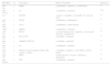
Lin et al. were the first researchers that observed that the expression profile of a subset of miRNAs could be used to classify high- and low-risk patients with a high sensitivity and specificity.21 To date, numerous miRNAs have been identified that regulate different oncogenic properties in NBs. In this section, we will highlight the miRNAs that have shown therapeutic effects in preclinical models (Table 1).
MicroRNAs with therapeutic potential in neuroblastoma.
| MicroRNA | ↑/↓ | Target genes | Effects of restoration | References |
|---|---|---|---|---|
| miR-34a | ↓ | TIMP2 | ↓ proliferation, ↑ apoptosis, ↓ vascularisation | 24 |
| miR-542-5p | ↓ | NA | ↓ proliferation, ↓ metastasis | 25,26 |
| Let-7 | ↓ | LIN28B | ↓ growth, ↓ clonability, ↓ cell viability, G1 cell cycle arrest | 28 |
| miR-27b | ↓ | PPARγ | ↓ proliferation, ↓ inflammation | 29 |
| miR-200a | ↓ | AP-2γ | ↓ proliferation, ↓ tumour growth | 31 |
| miR-9 | ↓ | MMP14 | ↓ proliferation, ↓ metastasis | 32 |
| miR-145 | ↓ | HIF-2α | ↓ cell and tumour growth, ↓ migration, ↓ invasion and metastasis, ↓ angiogenesis | 33 |
| miR-335 | ↓ | SOX4, TNC | ↓ clonability, ↓ metastasis | 34 |
| miR-363 | ↓ | AMDM15, MYO1B | ↓ clonability, ↓ metastasis | 34 |
| miR-183 | ↓ | NA | ↓ proliferation, ↑ apoptosis | 35 |
| miR-138 | ↓ | Proteins involved in apoptosis (Bcl-2, Bax, caspase-3, calpain…) | ↓ clonability, ↑ apoptosis, ↓ cell viability, ↓ tumour growth | 36 |
| miR-558 | ↑ | HPSE | ↓ tumour growth, ↓ invasion, ↓ metastasis, ↓ angiogenesis | 38 |
| miR-380-5p | ↑ | TP53 | ↓ proliferation, ↑ apoptosis | 40 |
NA, not analysed; ↑, increased; ↓, decreased.
The first miRNA considered to be a suppressor of NB was miR-34a.22 This miRNA is located in chromosomal region 1p36 (which is frequently deleted in NB) and regulated by the tumour suppressor gene TP53.23 Therefore, the therapeutic strategy is aimed at restoring the levels of this miRNA. In fact, its overexpression results in decreased cell proliferation and increased cell death by apoptosis, both in vitro and in vivo.24
One of the first strategies used to identify miRNAs with therapeutic potential was analysing the expression of different miRNAs and its correlation with different parameters of tumour aggressiveness. This approach led to the identification of miR-542, which seems to be one of the miRNAs with the lowest levels of expression in NB and is highly associated with poor patient survival. The overexpression of miR-542-5p in orthotopic models of NB caused a clear reduction in tumour volume.25 In a recent study on mice with NB xenotransplants, treatment with nanoparticles loaded with miR-542-3p resulted in decreased proliferation and increased cell death by apoptosis in the tumours.26
Another approach has been to study miRNAs that may play a role in the modulation of genes or processes known to be involved in NB. The first obvious focus of investigation was miRNA regulation of MYCN. The results of this research allowed the identification of a set of miRNAs that could suppress expression of the MYCN protein, including miR-34a/c, miR-449, miR-19ab, miR-101 and let-7/miR-202.27 However, there is only evidence in support of the therapeutic potential of let-7. Molenaar et al. elegantly demonstrated that LIN28B, a regulator of the processing of let-7, is amplified in high-risk NB and is therefore partly responsible for the low expression of let-7 and the ensuing overexpression of MYCN.28
The transcription factor PPARγ is also overexpressed in NB, which is partly due to the loss of expression of miRNAs such as miR-27b. The expression levels of miR-27b are low in NB, and treatment of mice with intraperitoneal injections of this miRNA resulted in a significant reduction of tumour growth.29 Treatment with miR-200a of mice with NB cell line xenotransplants achieved similar results. This miRNA belongs to the miR-200 family, the expression of which has been found to be altered in multiple tumours.30 The therapeutic effects of the increased expression of miR-200a have been attributed to inhibition of the expression of the transcription factor AP2-γ.31
Several studies have demonstrated that treatment with miRNAs may contribute to limiting the metastatic potential of NB cells. We find an early example of this in the work of Zhang et al., who analysed the regulation of the MMP-14 metalloproteinase by miRNAs. MMP-14 is a protein involved in migration, invasion and metastasis, so it can be a therapeutic target in NB. The authors demonstrated that overexpression of miR-9 can reduce the levels of MMP-14, leading to a reduction in tumour growth and metastasis of NB cells.32 The expression of MMP-14 is directly regulated by hypoxia-inducible factor 2 alpha (HIF-2α). Recent evidence shows that this factor can also be expressed in non-hypoxic tumour regions. In turn, this factor may be regulated epigenetically by miR-145. Overexpression of miR-145 in NB xenotransplants caused a reduction in the growth, angiogenesis and metastasis of NB cells.33
Another miRNA involved in metastasis is miR-335. The expression of this miRNA is directly suppressed by oncogene MYCN. This can lead to increased expression of several elements in the TGFβ signalling pathway (such as ROCK, MAPK1 and LRGR1), increasing the metastatic potential of NB cells. An unrelated study identified miR-335 and miR-363 as genes regulated by the gastrin-releasing peptide receptor (GRP-R) protein, involved in the tumorigenesis and metastasis of NB. The overexpression of miR-335 and miR-363 in NB cell lines reduced tumorigenesis and metastasis in vivo.34 It has also been observed that MYCN can cooperate with HDAC2 to repress expression of miRNA, specifically miR-183. Overexpression of miR-183 also sufficed to reduce tumour growth in vivo.35
In some instances, recovering the expression of a single miRNA may not be sufficient to achieve a therapeutic effect, but have beneficial effects in combination with conventional or experimental treatments. Such is the case of miR-138, which regulates the expression of the human telomerase reverse transcriptase (hTERT) gene, responsible for the indefinite growth of tumours. The increased expression of miR-138 achieved by injecting DNA in NB xenografts did not in itself affect tumour growth, but it potentiated the therapeutic effects of flavonoids such as apigenin.36
MicroRNAs that function as oncogenesResearch has identified several miRNAs that are overexpressed in tumours with poor prognosis that may function as oncogenes and whose inhibition may achieve therapeutic effects. One example is the recently described miR-558. This miRNA has an unconventional mechanism of action. Unlike the vast majority of miRNAs, whose main function is to inhibit translation and induce degradation of mRNA, miR-558 binds the promoter region of its target genes, such as the heparanase gene (HSPE), stabilising the mRNA and resulting in higher protein levels. This enzyme is involved in invasive processes, tumour growth and angiogenesis (reviewed in Ref. 37). On the other hand, Qu et al. demonstrated that intravenous injection of molecules for inhibiting the function of miR-558 (anti-miRs) resulted in decreased HSPE protein levels, which in turn led to decreased tumour growth, angiogenesis and lung metastases.38
One of the main miRNAs whose inhibition has great therapeutic potential is miR-380-5p. This miRNA was identified as a regulator of tumour suppressor gene TP53. While mutations or deletions of this gene are frequent in many tumours, changes in this gene are infrequent in NB.39 Nevertheless, we know that the function of TP53 is essential for the signal transduction of genotoxic stimuli such as those generated by chemotherapy drugs. Thus, one of the mechanisms by which tumour cells can tolerate the expression of the TP53 gene is by post-transcriptional control by means of miRNAs. This seems a plausible mechanism in a specific subset of NB patients. Specifically, high levels of miR-380-5p have been found in patients with MYCN-amplified disease and poor prognoses. Inhibition of miR-380-5p by intraperitoneal injection of anti-miRs has been shown to successfully reduce tumour growth in MYCN-dependent tumours by inducing TP53-mediated cell death.40
Conclusions and future perspectivesThe use of miRNAs in clinical practice is already a reality. In the field of oncology, the first molecule that has entered clinical testing is MRX34. This compound mimics miR-34, a miRNA that functions as a tumour suppressor whose expression is reduced in multiple tumours.
One limitation of these compounds is their administration by the intravenous route and devoid of any encapsulation, so that their distribution concentrates mostly in the liver, where they are very effective but are also metabolised and excreted rapidly, limiting their therapeutic effect in less perfused tissues. Therefore, more research is needed to improve the biodistribution of miRNAs, possibly by their encapsulation in nanoparticle vesicles manufactured with biocompatible materials, which result to greater stability and longer circulation in the bloodstream, allowing more time for their accumulation in tumour tissues and thus enhancing their antitumour efficacy.
FundingThis study has been funded by the Instituto de Salud Carlos III (CP11/00052, PI14/00561, RD12/0036/0016) and co-funded by the European Regional Development Fund (ERDF), Generalitat de Catalunya (2014-SGR-660) and the Marie Curie Career Integration Grants (n° 618301).
Conflict of interestsThe authors have no conflict of interests to declare.
Please cite this article as: Boloix A, París-Coderch L, Soriano A, Roma J, Gallego S, Sánchez de Toledo J, et al. Nuevas estrategias terapéuticas para el neuroblastoma basadas en el uso de microRNAs. An Pediatr (Barc). 2016;85:109.e1–109.e6.
