Journal Information
Images in Paediatrics
Full text access
Available online 5 April 2024
Phantom bone disease
Enfermedad del hueso fantasma
Visits
60
Gerardo Rivera-Silvaa,
Corresponding author
gerardo.rivera@udem.edu

Corresponding author.
, Hugo C. Mora-Magallónb, Miguel A. Guerrero-Ferreirac, Samantha Moriel de Leóna
a Departamento de Ciencias Básica, Escuela de Medicina, Universidad de Monterrey, Monterrey, NL, México
b Servicio de Odontología Pediátrica, Hospital Dr. José Torres Orozco”, Morelia, Mich. México
c Facultad de Odontología, Universidad Michoacana, Morelia, Mich. México
This item has received
Received 07 September 2021. Accepted 01 October 2021
Article information
Full Text
Bibliography
Download PDF
Statistics
Figures (2)
Full Text

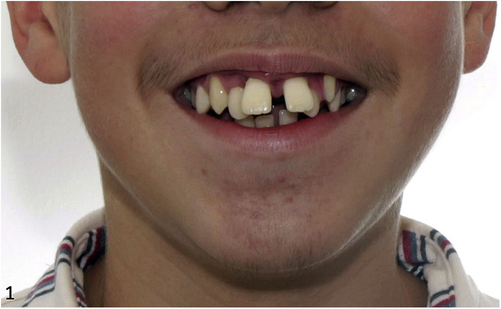
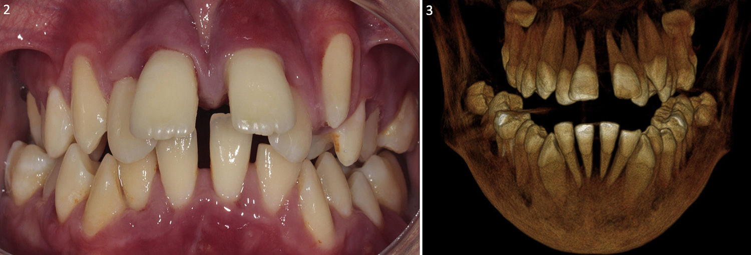
We present the case of a boy aged 12 years who presented with loose teeth that had erupted 6 years earlier. There was no history of trauma, infection or cancer involving the upper or lower jaw. The oral examination found an adequate mouth opening (Fig. 1), with small displacement of the teeth on palpation, and no signs of bone development, the teeth were simply covered by a reddened gum (Figure2 Fig. 2 ). The rest of the physical examination and the blood tests were normal. The 3D dental cone CT scan evinced the presence of osteolytic lesions in the mandible and absence of alveolar bone in the teeth of both the maxilla and the mandible, giving them the appearance of floating teeth (Figs. 2 and 3). The patient received a diagnosis of Gorham-Stout syndrome. It was managed with several bone grafts to correct the bone defects.

Figure 1.

General appearance of the mouth opening of the patient.

(0.06MB).
Figure 2 and 3.

Frontal view of the dentition of the patient, showing “floating” teeth associated with erythematous gums. Three-dimensional CT images showing a lack of alveolar bone in all teeth and osteolytic lesions in the mandible.

(0.1MB).
Commentary

Gorham-Stout syndrome, also known as phantom bone disease, is a disease of unknown aetiology characterised by osteolytic lesions that is frequently associated with benign vascular or lymphatic proliferation.1,2 It may affect any bone, but involves the skull, mandible and shoulder most frequently. In some cases, it may cause pain or swelling secondary to a fracture. It is a diagnosis of exclusion, since it is a rare disease and laboratory tests are usually normal, only imaging findings are abnormal.2 There is also substantial variation in its management, which is personalised and based on steroids, bisphosphonates, radiation therapy or surgery for transplantation of bone grafts. However, cases of spontaneous remission have also been reported.3

Funding

This research did not receive any external funding.

References
[1]
S.M. Gondivkar, A.R. Gadbail.
Gorham-Stout syndrome: a rare clinical entity and review of literature.
Oral Surg Oral Med Oral Pathol Oral Radiol Endod., 109 (2010), pp. e41-8
[2]
D.V. Patel.
Gorham’s disease or massive osteolysis.
Clin Med Res., 3 (2005), pp. 65-74
[3]
G. Gutiérrez Schiaffino, I. Leiva Gea, B. Martín Tejedor, J.M. Jiménez Hinojosa, A. Madrid Rodríguez, A. Urda Cardona.
¿Es la actitud expectante en el síndrome de Gorham una opción terapéutica?.
Anales de Pediatría., 81 (2014), pp. e64-5
Idiomas
Anales de Pediatría (English Edition)
Article options
Tools
es en

¿Es usted profesional sanitario apto para prescribir o dispensar medicamentos?

Are you a health professional able to prescribe or dispense drugs?