

We present the case of a primigravida, 33 years of age, with no medical or surgical history of interest. Ultrasound examination at 28 weeks of gestation confirmed the presence of a 28mm×17mm mass in the umbilical cord, with an umbilical cord diameter of 16mm, and a small anechoic area with thin walls suggestive of hernial oedema.
The patient had a normal delivery at 40+1 weeks of gestation, giving birth to a girl that weighed 3290g and had an Apgar score of 9/10.
At birth, we observed an umbilical cord with a 4.5cm×2cm×1.8cm bulge protruding from its normal insertion site at the abdomen, lined with amniotic membrane through which could be seen a firm, wine-red mass located 1cm away from the navel that was irreducible, with no accompanying symptoms (Fig. 1). Based on the examination findings, we considered the differential diagnosis of abdominal wall defect and umbilical cord mass.

The surgery involved the opening of the amniotic membrane in layers, revealing a solid mass in direct contact with the umbilical vein and with an intraperitoneal communication with the round ligament of the liver. The vascular structures and remnants of the umbilical cord were ligated, the mass fully resected, and the umbilical defect closed. There were no postoperative complications and the patient was discharged 5 days after the surgery.
The mass was submitted to the anatomical pathology department for investigation, and gross examination showed a well-defined brownish nodule measuring 2.5cm, with a microgranular appearance upon sectioning that corresponded to hepatic tissue with preserved architecture at the histological level. The tissue surrounded a cyst-like structure consisting of gallbladder wall tissue that was compatible with a hepatobiliary choristoma.
Ectopic liver is a rare condition described as the presence of hepatic tissue outside the liver and with no hepatic connection.1
The literature has reported the gallbladder as the most common location of ectopic liver, and it can also be found in the thorax, pancreas, spleen, hepatic ligaments, pylorus, greater omentum, oesophagus, gastric mucosa, adrenal cortex, retroperitoneum, pericardium, placenta and umbilical cord.
Several theories attempt to explain the appearance of ectopic liver in locations other than the gallbladder, such as the development of an accessory lobe that loses its connection with the main liver body, the migration of part of the pars hepatica to other sites where ectopic tissue then develops, or the trapping of hepatocytes by the adjacent mesenchyma during the formation of the liver sinusoids and their subsequent migration to more distant regions, such as the umbilical cord, while the connection with the main liver may be maintained through the umbilical vein.
The differential diagnosis of umbilical cord masses is complex and must include cyst and pseudocyst, haematoma, umbilical artery aneurysm, haemangioma, teratoma, angiomyomyxoma, patent urachus, ectopic liver, as well as the most common diseases of the umbilical cord, which are umbilical cord hernia, gastroschisis and omphalocoele.
Ectopic liver in the newborn is usually diagnosed by chance following imaging tests or surgical procedures performed for unrelated reasons. However, it may be diagnosed due to complications like torsion, which manifests with abdominal pain, gastric outlet obstruction and respiratory distress syndrome, caused by the presence of hepatic tissue in supradiaphragmatic locations.
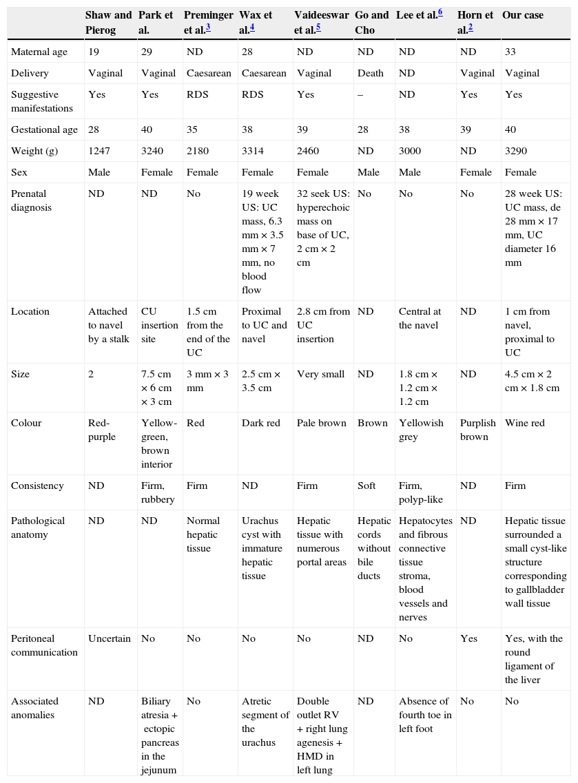
Only eight other cases of hepatic tissue in the umbilical cord have been described in the literature2–6 (Table 1), and the diagnosis of the umbilical cord mass was made prenatally in three of the nine cases, with the definitive diagnosis being made by anatomical pathology. On rare occasions it can be accompanied by symptoms of infection and be associated with other abnormalities, such as utrachal or biliary atresia, ectopic pancreas and heart and lung malformations. In our case, as happened in the one described by Horn et al.,2 we observed an intraperitoneal connection with the liver that may correspond to the round ligament, a vestige of the left umbilical vein.
Literature review of published hepatic choristoma cases.
| Shaw and Pierog | Park et al. | Preminger et al.3 | Wax et al.4 | Vaideeswar et al.5 | Go and Cho | Lee et al.6 | Horn et al.2 | Our case | |
|---|---|---|---|---|---|---|---|---|---|
| Maternal age | 19 | 29 | ND | 28 | ND | ND | ND | ND | 33 |
| Delivery | Vaginal | Vaginal | Caesarean | Caesarean | Vaginal | Death | ND | Vaginal | Vaginal |
| Suggestive manifestations | Yes | Yes | RDS | RDS | Yes | – | ND | Yes | Yes |
| Gestational age | 28 | 40 | 35 | 38 | 39 | 28 | 38 | 39 | 40 |
| Weight (g) | 1247 | 3240 | 2180 | 3314 | 2460 | ND | 3000 | ND | 3290 |
| Sex | Male | Female | Female | Female | Female | Male | Male | Female | Female |
| Prenatal diagnosis | ND | ND | No | 19 week US: UC mass, 6.3mm×3.5mm×7mm, no blood flow | 32 seek US: hyperechoic mass on base of UC, 2cm×2cm | No | No | No | 28 week US: UC mass, de 28mm×17mm, UC diameter 16mm |
| Location | Attached to navel by a stalk | CU insertion site | 1.5cm from the end of the UC | Proximal to UC and navel | 2.8cm from UC insertion | ND | Central at the navel | ND | 1cm from navel, proximal to UC |
| Size | 2 | 7.5cm×6cm×3cm | 3mm×3mm | 2.5cm×3.5cm | Very small | ND | 1.8cm×1.2cm×1.2cm | ND | 4.5cm×2cm×1.8cm |
| Colour | Red-purple | Yellow-green, brown interior | Red | Dark red | Pale brown | Brown | Yellowish grey | Purplish brown | Wine red |
| Consistency | ND | Firm, rubbery | Firm | ND | Firm | Soft | Firm, polyp-like | ND | Firm |
| Pathological anatomy | ND | ND | Normal hepatic tissue | Urachus cyst with immature hepatic tissue | Hepatic tissue with numerous portal areas | Hepatic cords without bile ducts | Hepatocytes and fibrous connective tissue stroma, blood vessels and nerves | ND | Hepatic tissue surrounded a small cyst-like structure corresponding to gallbladder wall tissue |
| Peritoneal communication | Uncertain | No | No | No | No | ND | No | Yes | Yes, with the round ligament of the liver |
| Associated anomalies | ND | Biliary atresia+ectopic pancreas in the jejunum | No | Atretic segment of the urachus | Double outlet RV +right lung agenesis+HMD in left lung | ND | Absence of fourth toe in left foot | No | No |
HMD, hyaline membrane disease; ND, not described; RDS, respiratory distress syndrome; RV, right ventricle; UC, umbilical cord; US, ultrasound.
To conclude, we would like to highlight that when ultrasound examination reveals a mass in the umbilical cord we should consider the possibility of rare conditions, like the one described here, along with more common diseases.
Doppler ultrasound of the mass can be helpful to this end, although as we mentioned above, in most cases the definitive diagnosis will be made postnatally. At any rate, the histological characteristics of the lesion should not change the obstetric approach in the absence of intestinal or vascular involvement in the foetus, and the surgical approach will depend on the suspected diagnosis after birth.
Please cite this article as: González Ruiz Y, Cotaina Gracia L, Ruiz de Temiño M, González Esgueda AJ, Delgado Alvira MR. Revisión de casos publicados de coristoma hepático. Diagnóstico diferencial de masas de cordón umbilical. An Pediatr (Barc). 2015;83:132–134.
