Journal Information
Vol. 100. Issue 4.
Pages e3-e5 (01 April 2024)
Vol. 100. Issue 4.
Pages e3-e5 (01 April 2024)
Images in Paediatrics
Full text access
Self limiting sternal tumors of childhood in a 7 months old infant
Tumor esternal autolimitado en una lactante de 7 meses
Visits
104
Gonzalo Fuente-Lucasa,
Corresponding author
gonzalo.fuente.gfl@gmail.com

Corresponding author.
, Marina Cecilia Planells-Alduvinb, Paloma Tallón-Guerolab, Pedro Alcalá-Minagorrea
a Servicio de Pediatría, Hospital General Univesitario de Alicante, Alicante, Spain
b Servicio de Radiología Pediátrica, Hospital General Universitario de Alicant, Alicante, Spain
This item has received
Article information
Full Text
Bibliography
Download PDF
Statistics
Figures (3)
Show moreShow less
Full Text

Self-limiting sternal tumours of childhood (SELSTOC) are characterised by the development of a sternal tumour unrelated to previous trauma, rapidly growing and with spontaneous resolution.1,2

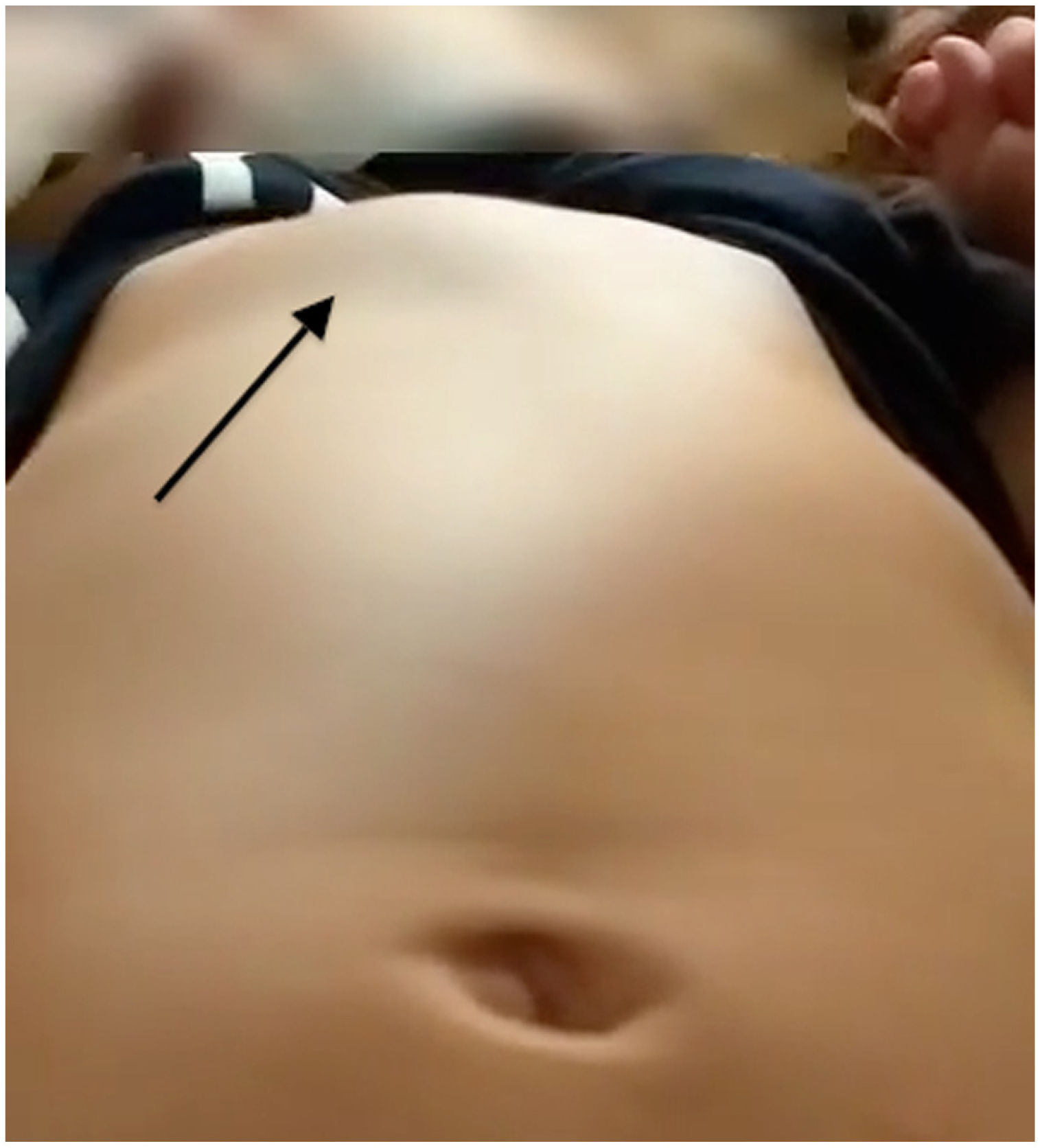
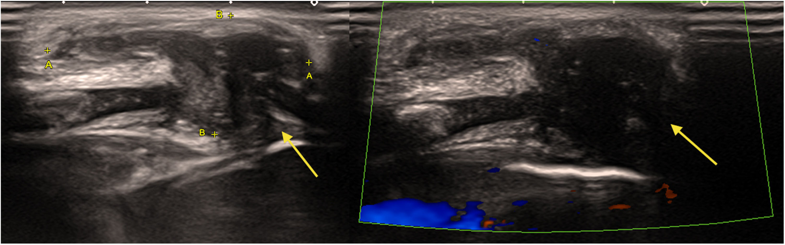
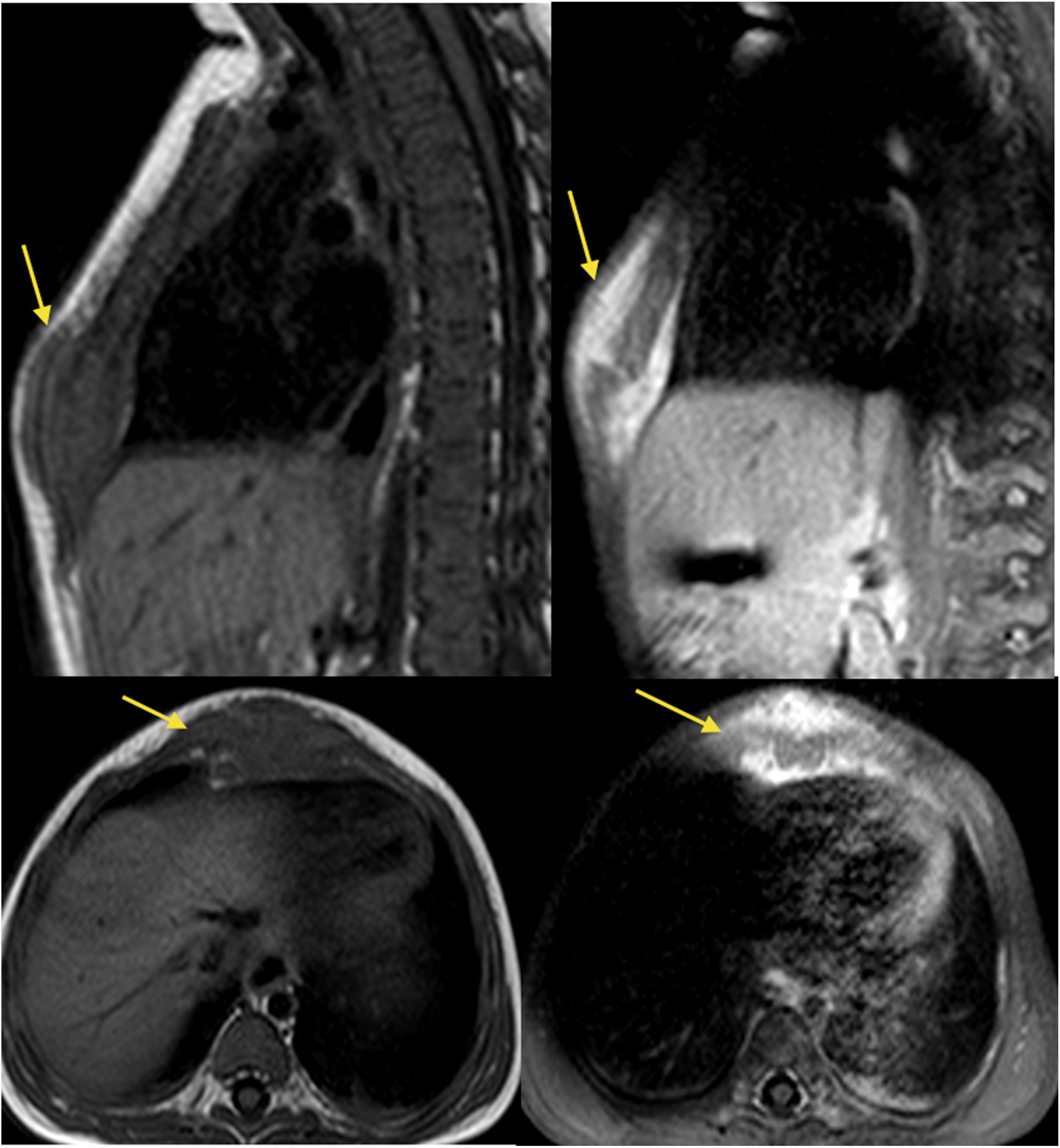
We present the case of a female infant aged 7 months who presented with a tumour in the lower third of the sternum (Fig. 1) detected 2 months prior, of solid consistency, mildly tender on palpation and coughing. The family reported there was no previous trauma and no associated constitutional symptoms. The ultrasound scan (Fig. 2) and magnetic resonance imaging (Fig. 3) showed a solid lesion (31 × 14 × 16 mm) at the level of the cartilage between the sternal body and the xiphoid process with involvement of the cartilage of the right 6th and 7th costochondral joints. The examination of a biopsy specimen of the lesion showed mature fibrocartilaginous material with no signs of malignancy. The patient remained in follow-up at the outpatient level, with observation of the gradual regression of the tumour until it nearly disappeared and resolution of symptoms.

Figure 1.

Tumour with a solid consistency in the lower third of the sternum (arrow).

(0.11MB).
Figure 2.

Soft tissue lesion in the caudal sternum measuring approximately 31 × 14 × 16 mm. The lesion seemed to extend to the sternal ossification centres and the cartilage of the right costochondral junctions, with low Doppler flow values.

(0.25MB).
Figure 3.

T1-weighted MRI, pre- and post-gadolinium: sagittal and axial views showing a lesion of the soft tissues surrounding the distal sternum with uptake of contrast.

(0.38MB).

The detection of a solid sternal mass in childhood requires differential diagnosis including inflammatory, infectious and malignant diseases. If a patient presents with a rapidly growing mass and no relevant history, the possibility of SELSTOC must be considered. This clinical spectrum could encompass Tietze syndrome, an infrequent self-limited inflammation of the costochondral junction described in the paediatric age group.3 In some cases, imaging tests may be required, and possibly a biopsy for the histological examination of the lesion. The course is self-limiting, usually with spontaneous regression, and the symptoms may be managed with analgesics as needed.

References
[1]
M.L. Winkel, M.H. Lequin, J.R. Bruyn, C.P. van de Ven, R.R. Krijger, R. Pieters, et al.
Self-limiting sternal tumors of childhood (SELSTOC).
Pediatr Blood Cancer., 55 (2010), pp. 81-84
[2]
D. Adri, T. Kreindel.
Tumores esternales autolimitados en edad pediátrica: a propósito de dos casos.
Radiología., 61 (2019), pp. 167-170
[3]
M. Mukamel, L. Kornreich, G. Horev, A. Zebaria, M. Mimouni.
Tietze’s síndrome in children and infants.
J Pediatr., 131 (1997), pp. 774-775
Idiomas
Anales de Pediatría (English Edition)
Article options
Tools
es en

¿Es usted profesional sanitario apto para prescribir o dispensar medicamentos?

Are you a health professional able to prescribe or dispense drugs?