


The Chiari network (CN) is a thin membrane attached to the right atrium resulting from the incomplete resorption of the right sinus venosus.1,2 Although it is generally benign, the persistence of this remnant has been found to be associated with cardiovascular complications such as supraventricular arrhythmias, interatrial septal defects, thromboembolism or cyanosis.2,3 Its diagnosis usually results from an incidental finding. The treatment is conservative on account of the spontaneous involution of the CN in the first months of life. However, some patients may require surgery.3
We present the cases of 4 neonates with cyanosis secondary to CN persistence that resulted in a right atrial flow pattern, directing blood flow from right to left through the patent foramen ovale (PFO) (Fig. 1A–D). One patient also developed self-limited supraventricular tachycardia, leading to initiation of oral treatment with flecainide. Three patients had favourable outcomes with resolution of cyanosis as the shunt through the PFO reversed. Another patient required surgery at 2 months of age due to persistent cyanosis and tricuspid valve obstruction caused by prolapse of the CN (Fig. 1E-F).
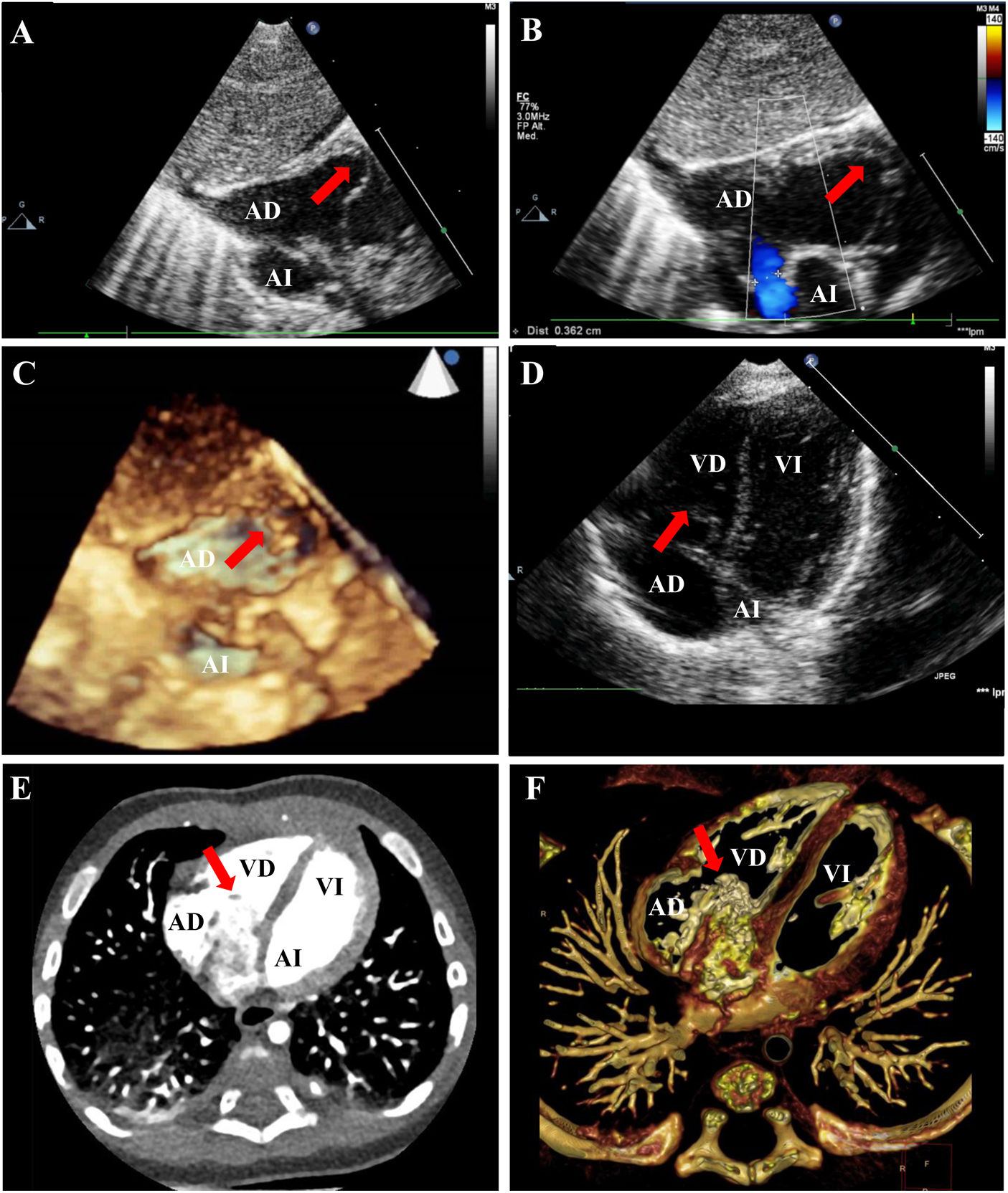
A. Echocardiogram, subcostal view, patient 1. B. Echocardiogram, subcostal view, patient 1. Right-to-left shunt through patent foramen ovale (blue flow). C. 3D echocardiogram, subcostal view, patient 1. D. Echocardiogram, 4-chamber view, patient 1. E-F. CT angiography, patient 4.
CT, computed tomography; LA, left atrium; LV, left ventricle; RA, right atrium; RV, right ventricle. Red arrow: Chiari network.
Although it is an apparently benign structure, persistence of the CN should be included in the differential diagnosis of cyanosis in newborns.
FundingThis research did not receive any external funding.
Conflicts of interestThe authors have no conflicts of interest to declare.
We thank Dr. Flavio Zuccarino for his collaboration in obtaining the computed tomography images.
