


Central nervous system (CNS) tumours are the most common solid tumours in children. Among these, the low-grade gliomas are the most common type, accounting for up to 30–50% of them.
Patients and methodsA retrospective analysis was carried out on the epidemiology, clinical characteristics, tumour location, histology, treatment, outcome and long-term sequelae of 111 patients diagnosed with low-grade glioma in the Niño Jesús Children's Hospital of Madrid from January 2002 to December 2011.
ResultsOf the 111 patients, there were 57 boys and 54 girls. The mean age was 7.26 years (range, 2 months–19 years). The most common symptoms of presentation were headache (27%) and vomiting (19%). The most common locations were the cerebral hemispheres (38%), followed by the brainstem (27.4%), and cerebellum (18.5%). Histological examination was performed in 89 patients (80.18%). Pilocytic astrocytoma was the most common histological type. Diagnostic biopsy was performed in 20 patients (22.5%), partial resection in 38 patients (42.7%), and total resection in 31 patients (34.8%). Sixteen patients received chemotherapy (14%), and eighteen patients received radiotherapy (16%). Overall survival was 88.3%. Long term hearing, visual and endocrine sequelae were noted in one, five, and four patients, respectively.
ConclusionsThe most common histological type is pilocytic astrocytoma. Overall survival was 88.3%. Only 9% of patients had some kind or auditory, visual or endocrine sequelae.
Los tumores del SNC son los tumores sólidos más frecuentes en la edad pediátrica. Dentro de ellos los gliomas de bajo grado constituyen el tipo más común de tumour del SNC en niños, representando hasta el 30–50% de los mismos.
Pacientes y métodosAnálisis retrospectivo de las características epidemiológicas, manifestaciones clínicas, localización del tumour, histología, tipo de tratamiento si lo ha recibido, evolución y secuelas a largo plazo de 111 pacientes diagnosticados de glioma de bajo grado en el Hospital Infantil Universitario Niño Jesús de Madrid entre enero de 2002 y diciembre de 2011.
ResultadosDe los 111 pacientes 57 eran niños y 54 niñas. La edad media fue de 7,26 años (intervalo 2 meses-19 años). Los síntomas de presentación más frecuentes fueron la cefalea (27%) y los vómitos (19%). Las localizaciones más frecuentes fueron los hemisferios cerebrales (38%), seguido del tronco cerebral (27,4%) y del cerebelo (18,5%). Se realizó estudio histológico en 89 pacientes (80,18%), siendo el astrocitoma pilocítico el tipo histológico más frecuente. Se realizó biopsia diagnóstica en 20 pacientes (22,5%), resección parcial en 38 pacientes (42,7%) y resección total en 31 pacientes (34,8%). Recibieron quimioterapia 16 pacientes (14%) y radioterapia 18 pacientes (16%). La supervivencia global fue del 88,3%. Un paciente presentó secuelas auditivas, 5 pacientes presentaron secuelas visuales y 4 pacientes secuelas endocrinas.
ConclusionesEl tipo histológico más frecuente es el astrocitoma pilocítico. La supervivencia global fue del 88,3%. Solo el 9% de los pacientes presentaron algún tipo de secuela auditiva, visual o endocrinológica.
Tumours of the central nervous system (CNS) are the most frequent solid tumours in the paediatric age group. Among them, gliomas are unquestionably the most common subtype, accounting for 46–70% of the patients in published case series.1
Gliomas are a heterogeneous group of tumours that are classified histologically into two groups, low-grade and high-grade, as specified by the World Health Organisation (WHO) criteria published in 2007.2 Low-grade gliomas are the most common type in the paediatric population, accounting for 30–50% of CNS tumours.3–6
In the WHO classification scheme, low-grade gliomas can be further classified as grade I (pilocytic astrocytoma) or grade II (fibrillary astrocytoma) tumours (Table 1).2 While the cerebellum is the most frequent location for gliomas, they can appear anywhere in the CNS. The symptoms vary according to location and usually appear months before the diagnosis. Surgery is the mainstay of treatment, and complete resection is the most important predictor of survival.
WHO low-grade glioma classification scheme.
| Astrocytoma |
| Grade I |
| Pilocytic astrocytoma |
| Subependymal giant cell astrocytoma |
| Grade II |
| Diffuse astrocytoma (fibrillary, protoplasmic, or gemistocytic) |
| Pilomyxoid astrocytoma |
| Pleomorphic xanthoastrocytoma |
| Oligodendroglial tumour |
| Grade II |
| Oligodendroglial tumour |
| Neuronal and mixed neuronal-glial tumours |
| Grade II |
| Ganglioglioma |
| Gangliocytoma |
| Desmoplastic infantile ganglioglioma |
| Dysembryoplastic neuroepithelial tumour |
In this review we have conducted a retrospective analysis of the cases of 111 paediatric patients with low-grade gliomas diagnosed at the Hospital Infantil Universitario Niño Jesús over a 10-year period.
Patients and methodsWe conducted a retrospective study of the patients diagnosed with low-grade glioma at the Paediatric Oncology Department of the Hospital Infantil Universitario Niño Jesús in Madrid over a period of 10 years, between January 1, 2002 and December 31, 2011.
For each patient, we collected data on demographic characteristics (age, sex), clinical manifestations, time to diagnosis, tumour location, histology, type of treatment received (if any), outcome, and long-term sequelae. The inclusion criteria for patients were the following: (a) age at diagnosis equal to or lower than 20 years; and (b) grade I or II glial tumour, as defined by the WHO classification, or low-grade glioma diagnosed on the basis of magnetic resonance imaging (MRI) findings. Gliomas were diagnosed as low-grade when MRI scans showed homogeneous intraaxial lesions that appeared hypointense in T1- and hyperintense in T2-weighted images, with minimal or absent post-contrast enhancement. Diffusion or spectroscopic MRI was performed in some patients, classifying gliomas as low-grade when they showed no resistance to diffusion and no alterations in N-acetylaspartate, choline and lipid peaks, respectively.7–9
ResultsDuring the 10 years of the study, a total of 111 patients were diagnosed with low-grade glioma in the Hospital Infantil Universitario Niño Jesús of Madrid. Of those 111 patients, 57 were boys (51.4%) and 54 girls (48.6%). The mean age at diagnosis was 7.26 years, with a standard deviation of 7 years (range, 0.16–18.66 years).
The most frequent presenting symptom was headache (27%), followed by vomiting (19%), epileptic seizures (18%), ataxia (16%) and cranial nerve involvement (14%). Three patients (2.7%) had onset with torticollis and one (0.9%) with visual and auditory hallucinations. Five of the patients presented with a papilloedema at diagnosis. The onset of symptoms had occurred more than one month prior to diagnosis in all patients, and in 37% it had occurred more than 6 months before. Of the 111 patients, 5 had been diagnosed with neurofibromatosis type 1, and 2 with tuberous sclerosis.
When it came to location, the most common site for the tumour was the brain (including the optic pathway and the hypothalamus) in 43 patients (38%), followed by the brainstem in 31 patients (27.4%), the cerebellum in 21 patients (18.5%), the cerebral ventricles in 7 patients (6.2%) and the spinal cord in 7 patients (6.2%). Only one patient, a 2-month-old infant with hypothalamic astrocytoma confirmed by histological examination, had leptomeningeal dissemination at the time of diagnosis.
The diagnosis was made by histological examination in 89 patients (80.18%) and based on MRI findings (as previously described) in the remaining 22 patients (19.82%). The histological diagnoses included 39 pilocytic astrocytomas (43.8%), 14 gangliogliomas (15.7%), 5 neuronal–glial tumours (5.6%), 2 diffuse astrocytomas (2.2%), 2 pilomyxoid astrocytomas (2.2%), 1 pleomorphic xanthoastrocytoma (1.1%), 1 subependymal giant cell astrocytoma (1.1%), and 25 tumours diagnosed as low-grade astrocytomas (28%).
Of the 22 patients in which it was not possible to perform a histological examination and who were diagnosed based on radiological findings, 16 had the tumour located in the brainstem, 3 in the optic nerve, 2 in the spinal cord, and 1 in a cerebral ventricle. The three patients with optic pathway gliomas, one of whom had bilateral involvement, were diagnosed with neurofibromatosis type 1, and the patient with a ventricular tumour was diagnosed with tuberous sclerosis with suspected subependymal giant cell astrocytoma.
In terms of treatment, 89 patients underwent surgery. In 20 of them (22.5%) it was only possible to perform a biopsy for the purpose of diagnosis. Of the remaining 69, 38 patients (42.7%) underwent a partial resection, and a complete resection could be done in 31 patients (34.8%). Chemotherapy was given to 16 patients (14.4%). Among them, seven had undergone a partial resection, a diagnostic biopsy alone had been done in four, and no surgery had been performed in the remaining five. When it came to the chemotherapy regimen, 10 patients were treated according to the latest SIOP protocol, one patient with a spinal cord glioma was treated with cisplatin and irinotecan, and the regimen used in the remaining 5 patients is unknown. Eighteen patients (16.2%) received radiotherapy, and their mean age was 6.82 years (range, 3.25–10.08 years). Seven of these patients had a brainstem tumour (4 of them confirmed by biopsy), and being younger than 8 years they were treated with radiation as a second-line treatment following chemotherapy. The remaining 11 patients were older at the time of radiotherapy, which was the first-line treatment. Of the 18 patients who received radiotherapy, 6 had undergone a partial resection and 5 a diagnostic biopsy. A patient diagnosed with chiasmatic pilocytic astrocytoma at 10 months of age was initially treated with chemotherapy, and later with radiation at 3 years of age after showing tumour progression. The patients treated with chemotherapy and/or radiotherapy had symptoms that varied according to tumour location in the brainstem, spinal cord, or on or near the optic pathway (cranial nerve paresis or paralysis and/or hemiparesis, or a decrease in visual acuity). None of the patients who underwent a complete resection were treated with radiation or chemotherapy at a later time.
The overall survival was 88.3%, with a median follow-up duration of 4.08 years (range, 0.04–9.72 years). Survival reached 100% in the 31 patients in which it was possible to perform a complete resection, and was 83.7% in the remaining 80 patients. Complete resection was associated with a higher overall survival (P<0.05). Of the 13 deceased patients, 5 had undergone a partial resection (none of them received radiation or chemotherapy), 2 only underwent a biopsy (both were treated with radiation and temozolomide) and surgical intervention had not been possible in any of the remaining six (one received radiotherapy alone, two received chemotherapy, and three temozolomide with radiotherapy). The main cause of death was tumour progression (Table 2).
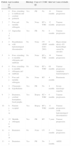
Characteristics of deceased patients.
| Patient | Age at Dx | Location | Histology | Type of Qx | CT/RT | Interval | Cause of death |
|---|---|---|---|---|---|---|---|
| 1 | 9y 1m | Pons, extending to midbrain and cerebral peduncle | Yes | PR | No | 14 months | Tumour progression |
| 2 | 7y | Pons and medulla oblongata | No | None | RT+TMZ | 12 months | Tumour progression |
| 3 | 13y 8m | Suprasellar | Yes | PR | No | 8 months | Tumour progression |
| 4 | 2m | Hypothalamic with leptomeningeal dissemination | No | None | CT (SIOP LGG 2001) | 6 months | Hypovolemic shock, (GI haemorrhage caused by steroids) |
| 5 | 6y | Pons, extending to medulla oblongata and midbrain | No | None | RT+TMZ | 18 months | Tumour progression |
| 6 | 4y 4m | Pons, extending to medulla oblongata and midbrain | No | None | RT+TMZ | 68 months | Tumour progression (anaplastic transformation?) |
| 7 | 5y | Pons and medulla oblongata | No | None | RT | 4 months | Pneumonia |
| 8 | 2m | Chiasmatic–hypothalamic | Yes | PR | No | 4 months | Severe neurologic sequelae |
| 9 | 3y | Brainstem, cerebral hemisphere, basal ganglia | Yes | Biopsy | RT+TMZ | 6 months | Tumour progression |
| 10 | 6y 3m | Parietal-occipital | Yes | Biopsy | RT+TMZ | 11 months | Tumour progression (with leptomeningeal dissemination) |
| 11 | 15y 8m | Medulla oblongata | Yes | PR | RT | 7 years | Tumour progression |
| 12 | 4y 8m | Brainstem | No | None | CT | 5 months | Tumour progression |
| 13 | 4y | Brainstem | Yes | PR | No | 2 months | ARDS |
Dx: diagnosis; Qx: surgery; CT: chemotherapy; RT: radiotherapy; y: years; m: months; PR: partial resection; TMZ: temozolomide; ARDS: acute respiratory distress syndrome; LGG: low-grade glioma; SIOP: Société Internationale d’Oncologie Pédiatrique.
There was tumour progression in 11 patients, of which only one had had a complete resection. Anaplastic transformation was suspected in one of them. This patient was a 4-year-old girl with a brainstem tumour that extended from the midbrain to the medulla oblongata diagnosed as a low-grade glioma based on MRI findings and which could not be treated with surgery. She received radiotherapy with concomitant temozolomide, which achieved tumour reduction and clinical improvement. The tumour remained stable until clinical and radiological data showed progression at 9 years of age. A histological examination was performed given this unusual evolution, confirming the presence of glioblastoma multiforme. Since a histological study had not been performed at diagnosis, the anaplastic transformation was an unconfirmed suspicion.
When it came to long-term sequelae, a patient with pontine astrocytoma (confirmed by histological examination) who had been treated with radiation developed mild hypoacusis; five patients with optic pathway glioma experienced a decrease in visual acuity (four of them had undergone chemotherapy and one radiotherapy); four patients with suprasellar or chiasmatic gliomas developed endocrine disorders (one developed hypothyroidism and diabetes insipidus, one panhypopituitarism, another precocious puberty, and the last one panhypopituitarism and diabetes insipidus).
DiscussionLow-grade gliomas usually have an indolent course, and there are even documented cases of spontaneous regression in children,10,11 contrary to what happens in adults, in whom these tumours are more aggressive. Due to their characteristic indolent behaviour, nearly 50% of patients have had clinical symptoms for 6 or more months by the time they are diagnosed.12 In our series, more than one month had elapsed from the onset of symptoms to diagnosis in every patient, and 6 or more months had elapsed in nearly 40% of patients.
The clinical manifestations develop as a function of the location and histology of the tumour, the age of the child, and the child's neural development. Most of them, including headache (usually in the morning), nausea, vomiting, decreased level of consciousness, papilloedema, and VI cranial nerve palsy, result from an increase in intracranial pressure caused by the obstruction of cerebrospinal fluid circulation. On the other hand, there are signs and symptoms that are associated with the tumour site. Thus, gliomas in the cerebellum may cause ataxia and dysmetria; gliomas in the cerebral hemisphere seizures, hemiparesis or changes in behaviour; gliomas in the hypothalamus and the pituitary gland, obesity, diabetes insipidus, endocrine disorders or visual abnormalities; optic pathway gliomas, visual abnormalities, proptosis or strabismus; brainstem gliomas, cranial nerve pair abnormalities (such as dysphagia or dysarthria) and long tract signs (hemiparesis, spasticity, hyperreflexia, Babinski's sign); and, last of all, cervicomedullary gliomas may cause torticollis, sensory deficits, or long tract signs and symptoms.3 On rare occasions, low-grade gliomas metastasise or progress to high-grade gliomas.3,13,14 Dissemination or metastasis has been described in 3–5% of patients at diagnosis, and in 7–10% of patients during the course of the disease, occurring more frequently in children younger than 1 year.14–19 The literature on the subject consists of single-case reports and a few small series, with the largest one comprising 13 patients.14,16–21 In our series, only one patient had leptomeningeal dissemination at the time of diagnosis, and anaplastic transformation was only suspected in one other. The first case corresponded to a 2-month-old boy with a parietal–occipital glioma confirmed by biopsy. The second case involved a patient with a brainstem tumour extending from the midbrain to the medulla oblongata diagnosed based on MRI findings without a histological examination. She was treated with radiotherapy with concomitant temozolomide, which achieved a reduction in tumour size and clinical improvement. The patient remained stable until her symptoms and radiological findings suggested tumour progression five years after diagnosis, at which time a biopsy was performed that confirmed a diagnosis of glioblastoma multiforme. Some authors have suggested that there is a link between radiotherapy and malignant transformation.22 On the other hand, according to a recent study, most pontine gliomas (up to 91%), especially those that are diffuse, are high-grade gliomas.23 Since no histological examination was performed in this patient at the time of diagnosis, the anaplastic transformation was only suspected. The long time interval that elapsed between diagnosis and tumour progression combined with the history of radiotherapy support the possibility of anaplastic transformation, while the tumour location at the brainstem with pontine involvement supports the possibility that she had a high-grade glioma from the beginning.
The literature up to date shows that the cerebellum is the most frequent site, with cerebellar low-grade gliomas accounting for 15–25% of all CNS tumours in children; followed by cerebral hemisphere gliomas (10–15%), gliomas of the deep midline structures (10–15%), optic pathway gliomas (5%) and brainstem gliomas (2–4%).4 In our series, the most common locations were the cerebral hemispheres (including the optical nerve) and the brainstem, unlike what is reported in the literature.
Children with neurofibromatosis type 1 account for the majority of gliomas of the optic pathway and the hypothalamus (over 70%).24 In fact, up to 15–20% of children with neurofibromatosis type 1 develop optic pathway gliomas, although only half of them develop symptoms and require treatment.25 In our series, out of the five patients who were diagnosed with neurofibromatosis type 1, three had low-grade optic nerve gliomas, with bilateral involvement in one.
Imaging techniques (computer tomography and preferably MRI) can be used to make an initial diagnosis, followed by a biopsy to confirm the presence of the tumour histologically. Craniospinal MRI can be used to explore the presence of leptomeningeal dissemination, which as noted above rarely occurs, but if it is suspected it should be performed to complement the cytological examination of the CSF.26 It is not always possible to do a biopsy, especially in tumours located in the optic nerve, the brainstem, or the deep brain structures. Thus, in order to preserve optic nerve function, a biopsy is not performed in children diagnosed with optic pathway or hypothalamic gliomas and imaging findings compatible with low-grade glioma, particularly in those with a neurofibromatosis type 1 diagnosis. Biopsies are only performed after careful consideration in cases of low-grade glioma of the brainstem or of the deep brain structures, especially if they are asymptomatic or show no progression in serial imaging studies.3 In our series, a biopsy was performed in only 7 of the 31 patients with suspected low-grade brainstem glioma, and the rest were diagnosed based on radiologic criteria as described above. Of the five patients with neurofibromatosis type 1, the three who had an optic pathway tumour suggestive of low-grade glioma did not undergo a biopsy; while a biopsy was performed in the remaining two (one with frontal hemisphere tumour and one with a spinal cord tumour).
Surgery is the mainstay of treatment. Complete resection of the tumour must be performed whenever possible, taking into consideration the sequelae that may result from it. Chemotherapy and/or radiotherapy are reserved for symptomatic patients in whom a complete resection could not be done. Since these are low-grade tumours, these treatments must only be done after careful consideration in patients that remain asymptomatic, as they do have side effects.27–30 Radiotherapy is associated with neuroendocrine and cognitive defects, vascular pathologies and second tumours, especially in patients with neurofibromatosis type 1.27–29 To avoid these complications, chemotherapy is usually the treatment of choice in younger children, as its side effects are fewer or less severe (such as carboplatin sensitivity).30 Most of the patients who received chemotherapy and/or radiotherapy in our series had symptoms associated with the location of the tumour in the brainstem, the optic pathway or surrounding areas, or the spinal cord (cranial nerve paresis or palsy and/or hemiparesis or decreased visual acuity), and radiotherapy was used as the first-line treatment in older children. It has been widely reported in the literature that the degree of surgical resection is the factor that shows the strongest association with overall and progression-free survival,12,31–35 with complete resection achieving survival rates of 90% or greater 10 years after diagnosis.12,26,31,33,36,37 In our series, there were no deaths among the patients in whom a complete resection could be done.
Tumour histology appears to be an independent predictor of progression. Thus, nonpilocytic tumour, and specifically diffuse fibrillary, histology is more highly associated with progression, recurrence, and anaplastic transformation, although as noted above the latter is rare in children.12,31,34,35,38 In our series, 11 patients had tumour progression, and anaplastic transformation was only suspected in one, as previously noted.
Survival rates are better in children than in adults, although the role of age as a prognostic factor is yet to be determined.1,26,35,38–40
In conclusion, survival in low-grade gliomas is high with currently available treatments, with an overall survival rate of 88.3% in our series, which rose to 100% in patients that underwent complete resection. On the other hand, survival with sequelae should not necessarily be considered a success, so we must minimise the sequelae associated with treatment, always taking into consideration the risk/benefit ratio to pursue a good quality of life. While the follow-up period was short in this case series, only 9% of the patients developed some kind of auditory, visual, or endocrine sequelae.
Conflicts of interestThe authors have no conflicts of interest to declare.
Please cite this article as: Pardal Souto M, Hernández Marqués C, Lassaletta Atienza A, Ruano D, Cormenzana M, Madero L. Gliomas de bajo grado: revisión de 10 años. Anales de Pediatría. 2015;82:68–74.
